Shoulder Pain Treatment in Noida - Causes, Diagnosis and When to See a Specialist

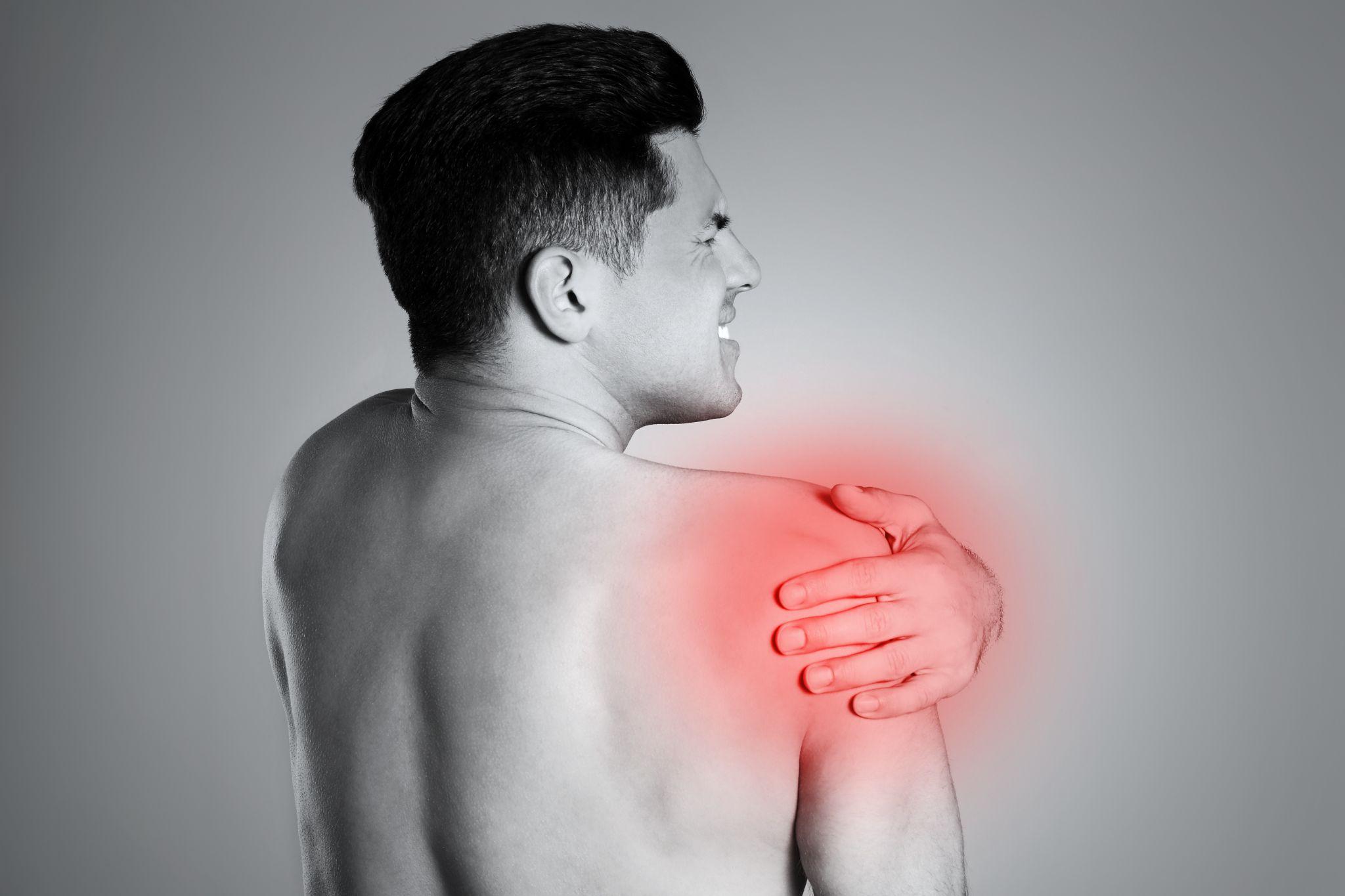
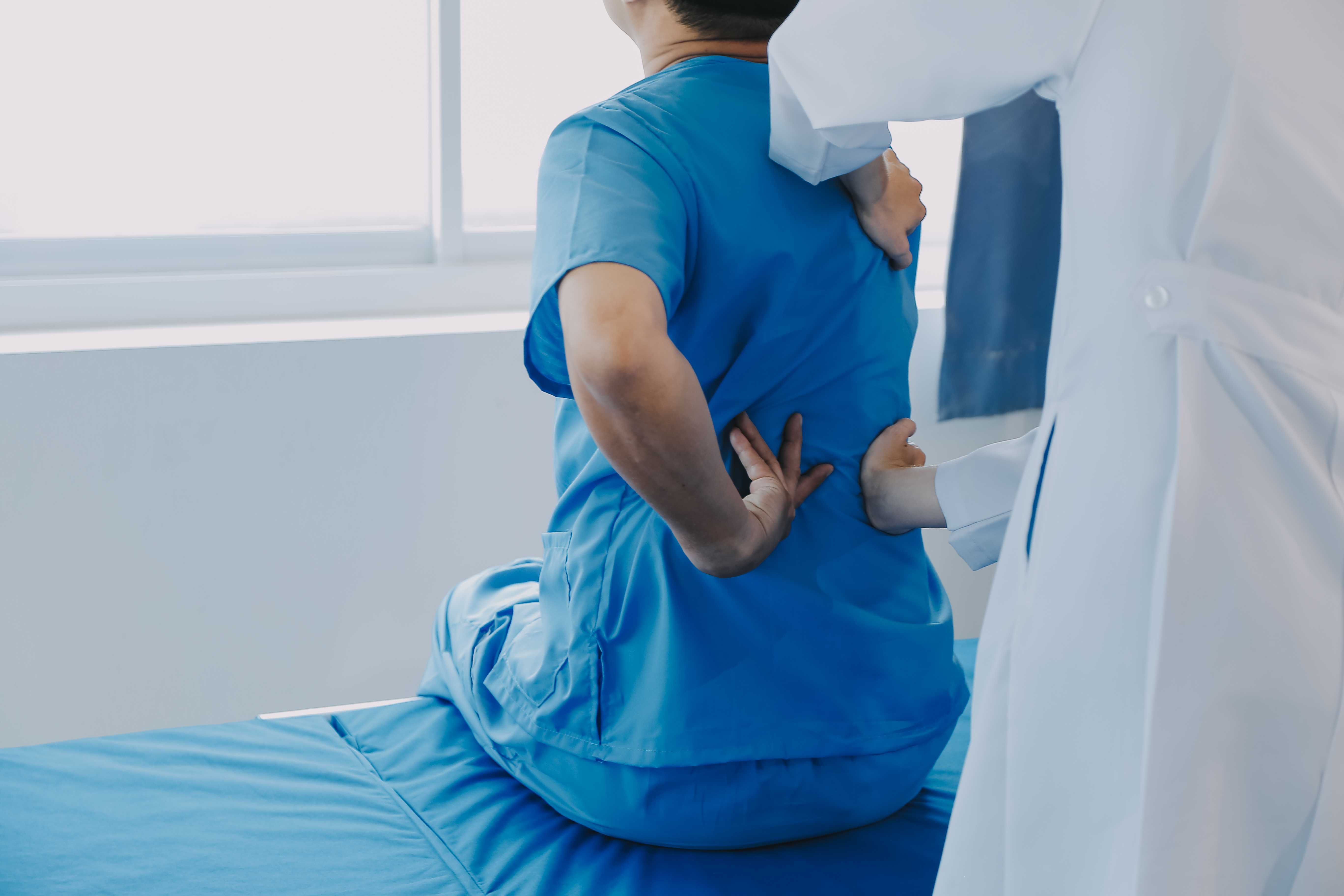
Person with rotator cuff pain.
The shoulder is the most mobile joint in the human body — capable of 360 degrees of movement in multiple planes. This extraordinary mobility comes at a cost: the shoulder sacrifices stability for range of motion, relying on a complex system of muscles, tendons, ligaments, and cartilage to keep the ball (humeral head) centred in the socket (glenoid) throughout its arc of movement.
When any component of this system fails — whether from acute injury, repetitive overuse, degenerative ageing, or inflammatory disease — the result is shoulder pain. And because so many different structures are involved, shoulder pain is one of the most diagnostically challenging symptoms in orthopedic practice.
In Noida, shoulder pain presentations span the full demographic range: frozen shoulder in a 45-year-old diabetic manager from Sector 62, rotator cuff tendinitis in a 35-year-old gym enthusiast from Noida Extension, shoulder OA in a 65-year-old from Sector 47, and an acute shoulder dislocation in a 22-year-old cricketer from Greater Noida. Each requires a completely different approach.
This blog maps the most common causes of shoulder pain in Noida's population, explains how they are diagnosed, and outlines what treatment is appropriate for each.
The Shoulder — A Brief Anatomy Reminder
Understanding shoulder pain requires knowing what might be causing it. The shoulder complex involves
Glenohumeral joint (GHJ): The ball-and-socket joint proper — the humeral head sitting in the glenoid socket of the shoulder blade (scapula). The socket is shallow and surrounded by the labrum — a cartilage ring that deepens it.
Rotator cuff: Four muscles (supraspinatus, infraspinatus, teres minor, subscapularis) and their tendons that surround the GHJ, stabilise it, and power most shoulder movements. The supraspinatus passes through the subacromial space — the gap between the top of the humerus and the acromion (the roof of the shoulder) — and is the tendon most commonly damaged.
Subacromial space: The space above the rotator cuff, below the acromion. Contains the subacromial bursa (a fluid-filled sac that reduces friction) and the supraspinatus tendon. When inflamed — from impingement, calcium deposits, or rotator cuff damage — this space is the source of most shoulder pain with overhead activities.
Acromioclavicular joint (ACJ): The joint at the top of the shoulder where the collarbone meets the acromion. A common site of arthritis and injury.
Biceps tendon: Attaches at the glenoid labrum (long head) and at the radial tuberosity in the elbow. Tendinitis and tears of the long head biceps are a common source of anterior shoulder pain.
Cervical spine: Not a shoulder structure, but a very important source of referred pain to the shoulder and arm. Cervical disc disease and facet arthritis commonly refer pain to the shoulder region — and must always be considered in the differential diagnosis of shoulder pain.
The Most Common Causes of Shoulder Pain in Noida
1. Subacromial Impingement and Rotator Cuff Tendinitis
The most common cause of shoulder pain overall in adults aged 30–60.
What it is: When the supraspinatus tendon becomes inflamed — from overuse, posture-related changes in the subacromial space geometry, or early degenerative change — it is "impinged" (compressed) between the humeral head and the acromion during overhead movement.
Who gets it: Office workers with poor posture (forward shoulder rounding narrows the subacromial space), gym enthusiasts with too much pressing and not enough pulling work (creates a muscle imbalance that raises the humeral head), overhead athletes (badminton, swimming, cricket fast bowlers), and anyone whose shoulder mechanics are altered by muscle weakness.
What it feels like:
- Pain at the front and outer side of the shoulder — over the deltoid
- Painful arc — pain between approximately 60 and 120 degrees of shoulder elevation (the range where the tendon is impinged); the arm can be raised fully with difficulty
- Night pain — characteristically worse when lying on the affected side
- Weakness with specific movements (pushing, reaching overhead)
Treatment:
- Physiotherapy (the cornerstone): rotator cuff and periscapular muscle strengthening, posture correction
- Subacromial corticosteroid injection: rapid and effective pain relief, allowing physiotherapy to progress
- Modification of aggravating activities
- Surgery (subacromial decompression) only when 3–6 months of conservative management have failed: arthroscopic removal of inflamed bursa and bone spur (acromioplasty), widening the subacromial space
2. Rotator Cuff Tears
Covered in detail in the rotator cuff tear blog. Key points
Partial tears: Conservative management first — physiotherapy, PRP for appropriate cases. Most partial tears respond without surgery.
Full-thickness tears in active patients: Arthroscopic rotator cuff repair produces excellent outcomes — but the recovery commitment (sling for 6 weeks, 4–6 months to full function) must be accepted.
Massive tears with rotator cuff arthropathy: Reverse shoulder replacement.
3. Frozen Shoulder (Adhesive Capsulitis)
Covered in detail in the frozen shoulder blog. Particularly common in Noida's diabetic population.
Key points: Three stages (freezing, frozen, thawing). Pain is the dominant problem in the freezing stage — corticosteroid injection and physiotherapy are most effective. Stiffness dominates the frozen stage — hydrodilatation and intensive physiotherapy. Arthroscopic capsular release for refractory cases.
Screening: A frozen shoulder in any patient should prompt a check of blood sugar (HbA1c) — frequently the first presentation of undiagnosed diabetes.
4. Biceps Tendinitis and SLAP Tears
Biceps tendinitis:
Inflammation of the long head biceps tendon where it passes through the bicipital groove on the humeral head. Produces anterior shoulder pain (pain at the front of the shoulder, along the bicipital groove). Worsens with:
- Lifting objects in front of the body
- Activities that require forearm supination (turning the palm upward)
- Pressing on the bicipital groove reproduces the pain
Treatment: NSAIDs, physiotherapy, biceps tendon sheath injection. In resistant cases, arthroscopic tenodesis (reattaching the tendon to the humerus in a better position).
SLAP Tears (Superior Labrum Anterior to Posterior):
Tears of the cartilage ring (labrum) at the top of the glenoid — where the long head biceps tendon attaches. Common in overhead athletes (cricket bowlers, swimmers, badminton players) and from acute traction injuries.
Produces a vague deep shoulder ache, clicking with certain movements, and pain with overhead activity. Diagnosed on MRI (with gadolinium contrast for full sensitivity). Managed arthroscopically — repair or debridement depending on tear pattern.
5. Shoulder Dislocation and Instability
Acute dislocation: Covered in the sports injuries blog. Requires immediate reduction and assessment of associated injuries (Bankart lesion, Hill-Sachs lesion, bony defects). Young athletes with first-time traumatic dislocation have very high recurrence rates without surgical stabilisation.
Chronic instability: A shoulder that subluxes (partially dislocates) or dislocates repeatedly. Produces apprehension — the sensation that the shoulder is about to come out. Arthroscopic Bankart repair produces 85–95% success in preventing recurrence.
Atraumatic instability: Some patients — particularly young, hypermobile individuals — have multidirectional shoulder instability without any significant trauma. Management is physiotherapy-focused (strengthening the dynamic stabilisers) rather than surgical in most cases.
6. Acromioclavicular Joint (ACJ) Problems
ACJ arthritis: A common cause of pain directly over the top of the shoulder — pressing on the joint or crossing the arm across the body (horizontal adduction) reproduces the pain. Responds to injection and physiotherapy. Arthroscopic ACJ excision arthroplasty for refractory cases.
ACJ injuries (shoulder separation): Ligaments connecting the collarbone to the acromion are torn — typically in falls onto the outstretched arm or directly onto the shoulder. Mild separations (Grade 1–2) heal conservatively. Severe separations (Grade 4–6) may require surgical reconstruction.
7. Shoulder Arthritis (Glenohumeral OA)
Less common than knee or hip OA but significantly disabling when present. Pain with movement, at rest, and at night. Grinding sensation with movement. Progressive restriction of rotation (external rotation is typically lost earliest, as in frozen shoulder).
Managed conservatively with physiotherapy and injections in earlier stages. Total or reverse shoulder replacement for advanced disease.
8. Calcific Tendinitis
Calcium deposits form within the rotator cuff tendons — most often the supraspinatus. When these deposits are in the "resorptive phase," they cause acutely severe shoulder pain — sometimes described as the worst pain the patient has ever experienced, requiring emergency presentation.
Diagnosis: Visible on plain X-ray. MRI provides more detail.
Treatment:
- Acute severe phase: subacromial steroid injection provides dramatic relief; occasionally, barbotage (ultrasound-guided needling of the calcium deposit) is performed
- Chronic calcium deposits: physiotherapy and injection. Extracorporeal shock wave therapy (ESWT) — acoustic wave treatment — has good evidence for refractory cases. Arthroscopic calcium removal for persistent symptomatic deposits.
Making the Diagnosis — The Assessment Process
When a patient presents to Dr. Mayank Chauhan with shoulder pain, the assessment follows a structured approach:
History:
- Onset and mechanism (acute injury vs. gradual onset)
- Location of pain (front, top, back, outer shoulder)
- Character (aching, sharp, burning, radiating down the arm)
- Aggravating movements (overhead, behind the back, lying on the side)
- Night pain
- Weakness
- Neurological symptoms (tingling or numbness — suggesting cervical spine rather than shoulder)
Examination:
- Active and passive range of motion — measured in all planes
- Strength testing of specific rotator cuff muscles
- Specific tests: Neer's impingement sign, Hawkins-Kennedy test (subacromial impingement), empty can test (supraspinatus), Speed's test (biceps tendon), O'Brien's test (AC joint and SLAP), apprehension test (instability)
- Cervical spine assessment — to exclude referred pain
Investigations:
- X-ray: First-line — shows glenohumeral OA, calcification, acromion morphology, ACJ arthritis, fractures
- Ultrasound: Dynamic assessment of rotator cuff tendons, bursae, biceps tendon. Good for guiding injections.
- MRI: Gold standard for soft tissue detail — rotator cuff tear extent and quality, labral pathology, biceps tendon, cartilage
Shoulder Pain Treatment at Prakash Hospital Noida
Dr. Mayank Chauhan, Senior Orthopedic Surgeon at Prakash Hospital, Sector 33, Noida, provides comprehensive shoulder assessment and treatment — from physiotherapy guidance and ultrasound-guided injections through to arthroscopic rotator cuff repair, Bankart stabilisation, SLAP repair, and shoulder replacement.
For patients in Noida and Greater Noida with shoulder pain that has persisted more than 4–6 weeks, or that is significantly limiting daily activities or sleep, a specialist shoulder consultation provides the specific diagnosis and targeted treatment plan.
Consultation hours: Monday to Saturday, 10 AM to 8 PM | Sunday, 10 AM to 2 PM.
To book a consultation, call the number listed on the website.
The Bottom Line
Shoulder pain is common, diverse in its causes, and reliably treatable when correctly diagnosed. The treatment that works for frozen shoulder is different from what works for rotator cuff tear — and what works for ACJ arthritis is different from what works for instability.
Accurate diagnosis — based on a thorough clinical assessment supplemented by appropriate imaging — is the foundation on which effective treatment is built.
To consult Dr. Mayank Chauhan, Senior Orthopedic Surgeon in Noida, call the number listed on the website.