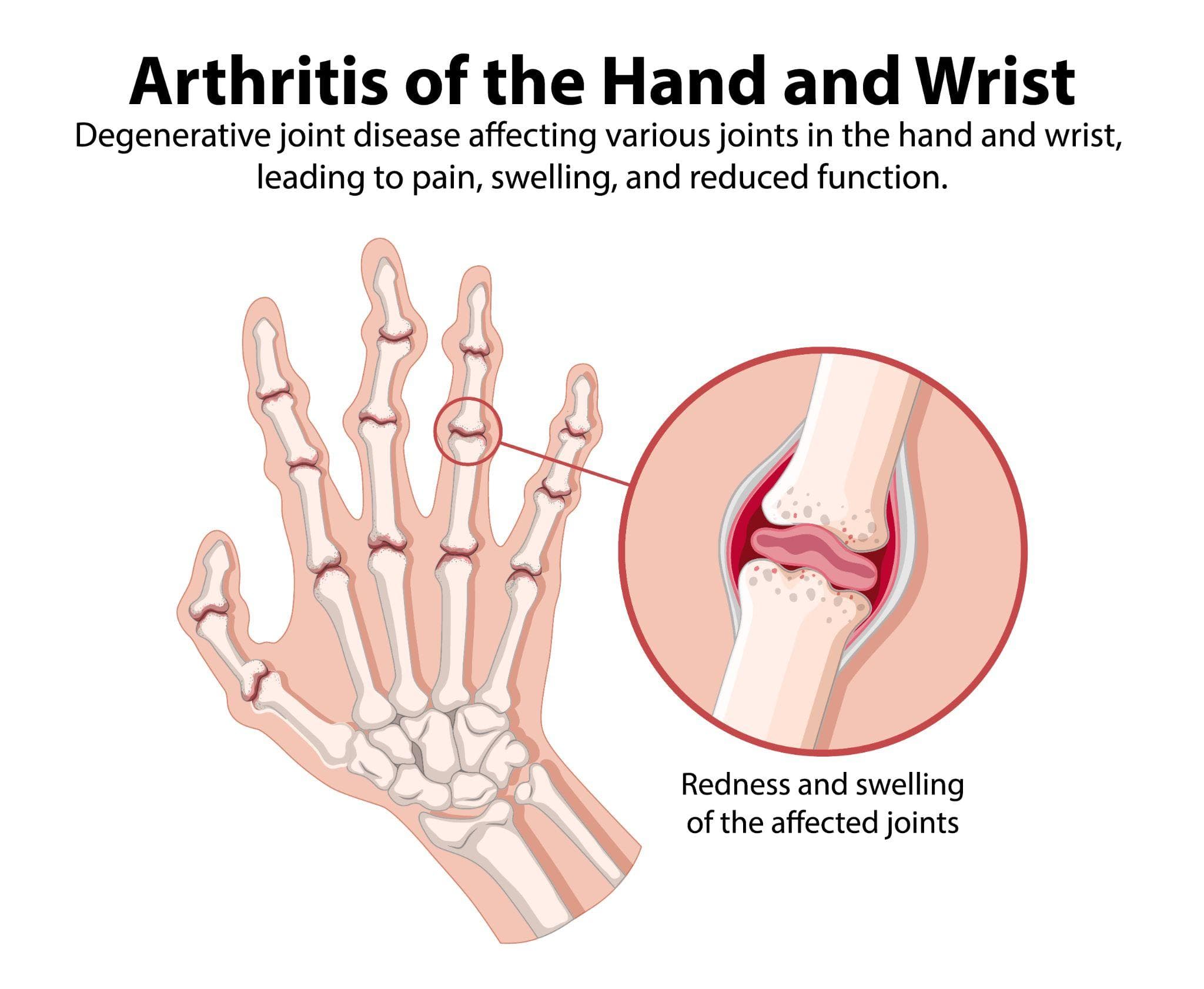
The 4 Stages Of Slipped Disc - What Your MRI Report Actually Means
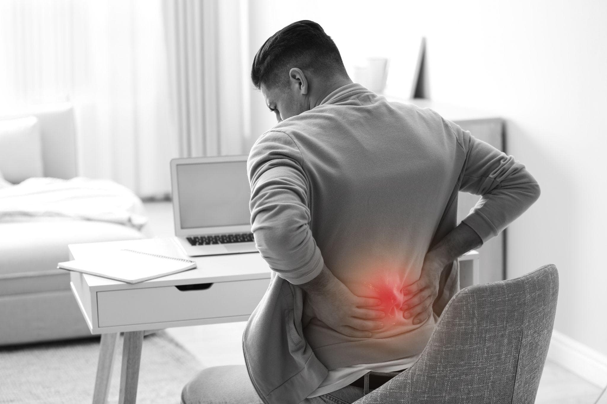
A man suffering from lower back pain while sitting, indicating slip disc due to poor posture.
If you've had an MRI of your lumbar spine in Noida, you've probably received a report with terms like "disc bulge at L4-L5," "disc herniation at L5-S1," or "disc extrusion with sequestration." And if you're like most patients, you read these words, felt a wave of anxiety, and weren't entirely sure what any of them meant.
Here's the key insight that will help you make sense of these reports: slipped disc is not one condition; it is a spectrum. What the radiologist is describing, whether it's a bulge, a prolapse, an extrusion, or a sequestration, tells you exactly how far along the spectrum the disc pathology is, and that directly affects the treatment.
This guide explains all four stages of disc herniation in plain language, what each one looks and feels like, and what treatment is appropriate at each stage.
Understanding The Disc First
Each intervertebral disc in the lumbar spine has two distinct components:
The annulus fibrosus: The tough outer ring made of concentric layers of fibrous collagen. Think of it as a reinforced tyre wall. It holds the disc together and contains the pressure of the inner material.
The nucleus pulposus: The soft, gel-like inner core of the disc. It contains water and proteoglycan molecules that give it its shock-absorbing properties. It is under considerable pressure within an intact disc.
When the annulus develops tears from degeneration, repeated stress, or sudden trauma, the nucleus can begin to push outward. The four stages of disc herniation describe how far this process has progressed.
Stage 1: Disc Bulge (Disc Protrusion Or Bulging Disc)
1. What It Is
In a disc bulge, the annulus fibrosus has begun to weaken, but it remains intact. The disc material doesn't push through the outer ring; instead, the disc simply extends (bulges) outward beyond its normal boundary at the disc level. The nucleus is under increased pressure but has not broken through. This is the earliest stage of the herniation spectrum. The disc is deformed but structurally contained.
2. What The MRI Report Says
"Disc bulge at L4-L5" or "posterior disc bulge with mild thecal sac effacement," the radiologist is describing that the disc is extending beyond its normal circumferential boundary without a focal tear.
3. Symptoms
Many disc bulges are asymptomatic and found incidentally when an MRI is done for another reason. When they are symptomatic:
- Dull lower back ache, often worse with prolonged sitting and better with movement.
- Stiffness, particularly in the morning.
- Mild discomfort with forward bending.
- Leg symptoms are uncommon unless the bulge is at a level that narrows the spinal canal significantly.
Important: A disc bulge on MRI does not automatically mean it is the cause of your symptoms. The clinical picture must match.
4. Treatment At Stage 1
Most disc bulges at Stage 1 are managed conservatively:
- Physiotherapy - core strengthening, McKenzie exercises, posture correction
- Activity modification - reducing positions that increase intradiscal pressure
- NSAIDs for pain management
- Ergonomic correction - particularly important for Noida's desk workers, where prolonged sitting is the primary driver.
Surgery is rarely indicated for a simple disc bulge without significant neurological compromise.
Stage 2: Disc Prolapse (Contained Herniation / Protrusion)
1. What It Is
In a disc prolapse, the inner nucleus has pushed through some of the inner layers of the annulus but is still contained by the outer layers. The disc material has moved beyond its normal position but has not yet broken through the outer ring of the disc. The disc is now under significantly greater stress at the tear site, and the prolapsed material can begin to exert pressure on nearby nerve roots.
2. What The MRI Report Says
"Disc herniation at L4-L5 with nerve root abutment" or "disc protrusion causing mild compression of the L5 nerve root," the radiologist is describing that the disc material is pressing toward or touching a nerve root.
3. Symptoms
This is where symptoms typically become more significant:
- Lower back pain, often worsening significantly.
- Radiating leg pain, the beginning of sciatica. Pain runs from the lower back into the buttock and down the leg, following the distribution of the compressed nerve root.
- Tingling or numbness along the nerve distribution.
- Pain is often worse with sitting, coughing, or sneezing (Valsalva test positive).
- Pain is often better with lying flat or gentle walking.
4. Treatment At Stage 2
Physiotherapy is the primary treatment, structured around the McKenzie method, core strengthening, and neural mobilisation:
- NSAIDs plus nerve pain medication (pregabalin) for leg symptoms.
- Epidural steroid injection if physiotherapy alone is insufficient after 4–6 weeks.
- Most patients at Stage 2 respond to conservative management within 6–12 weeks.
- Surgery (microdiscectomy) is considered if conservative treatment fails after 6–8 weeks, or if neurological weakness develops.
Stage 3: Disc Extrusion
1. What It Is
In disc extrusion, the nucleus pulposus has broken through the outer layers of the annulus fibrosus. The disc material has been extruded and pushed through, but remains connected to the parent disc. This is a more advanced herniation. The extruded material is now in direct contact with, or significantly compressing, the nerve root or the dural sac (the membrane containing the spinal cord and nerve roots).
2. What The MRI Report Says
"Disc extrusion at L5-S1 causing significant compression of the S1 nerve root" or "extruded disc fragment at L4-L5 with moderate spinal canal compromise."
3. Symptoms

A man holding his lower back due to lumbar pain, representing spine-related problems such as muscle strain or sciatica.
Extrusion typically produces more pronounced symptoms than prolapse:
- Severe sciatica, intense shooting or burning pain down the leg, often radiating to the calf and foot.
- Significant numbness and tingling along the nerve distribution.
- Possible motor weakness, difficulty lifting the foot (dorsiflexion weakness from L4-L5 extrusion), or weakness pushing down the foot (from L5-S1 S1 root involvement).
- Reduced or absent reflexes (knee jerk or ankle jerk, depending on level).
An important paradox: Very large extruded disc fragments, particularly if they are subligamentous or sequestrated, are actually among the most likely to spontaneously resorb. The immune response to a significant extrusion is more robust than to a contained bulge, and there are documented cases of large extrusions resolving completely on MRI over 6–12 months.
Treatment At Stage 3
Conservative treatment is still attempted first, but the threshold for escalation is lower:
- Aggressive physiotherapy plus epidural steroid injection.
- Surgical consultation is appropriate if there is motor weakness, if pain is severe and not responding to injection therapy, or if the clinical trajectory is worsening.
- Microdiscectomy is highly effective at this stage, the extruded fragment is directly accessible arthroscopically, and symptom relief is often immediate after surgery.
Stage 4: Disc Sequestration (Free Fragment)
1. What It Is
In disc sequestration, a fragment of the nucleus pulposus has completely separated from the parent disc and is now free within the spinal canal. This free fragment, sometimes called a "sequestered fragment," can migrate superiorly or inferiorly within the canal, moving away from the disc level itself. This is the most advanced stage of disc herniation. The fragment is no longer attached and cannot be resorbed by pulling back into the disc; it is a separate piece of tissue within the spinal canal.
What The MRI Report Says
"Sequestered disc fragment at L4-L5 level with severe compression of the L5 nerve root" or "extruded and sequestrated disc material at L5-S1 causing complete nerve root compression."
Symptoms
Sequestration can produce either very severe symptoms or, paradoxically, can be associated with sudden improvement:
- Severe symptoms (more common): Intense sciatica, significant motor weakness, possible bowel or bladder symptoms if the fragment is large enough to compromise the cauda equina.
- Paradoxical improvement: When a sequestered fragment migrates away from the nerve it was compressing, symptoms can suddenly improve significantly. This does not mean the problem has resolved; the fragment remains in the canal.
- Cauda Equina Syndrome Alert: A large sequestrated fragment at the L4-L5 or L5-S1 level can compress the cauda equina nerve bundle, causing loss of bladder or bowel control. This is a surgical emergency. If loss of bladder or bowel function occurs in a patient known to have a disc herniation, emergency surgical evaluation is required within hours.
Treatment At Stage 4
- If neurological deficit is progressing or if cauda equina syndrome is present: urgent surgery
- If symptoms are stable and conservative treatment is being tried, a shorter window before surgery is appropriate.
- Microdiscectomy at this stage involves both removing the sequestered fragment and the remaining disc material at the source level.
Reading Your MRI Report - A Quick Reference
| MRI Term | Stage | What It Means | Treatment Priority |
|---|---|---|---|
| Disc bulge / protrusion | Stage 1 | Contained, no tear, disc extending slightly | Conservative |
| Disc herniation / prolapse | Stage 2 | Annular tear, nucleus pushing through inner layers but contained | Conservative + injection if needed |
| Disc extrusion | Stage 3 | Nucleus through all annular layers, still attached to disc | Conservative first, lower surgical threshold |
| Sequestrated / free fragment | Stage 4 | Fragment completely separated, in spinal canal | Surgical if neurologically compromised |
What Stage Means For Prognosis
A common anxiety: patients with Stage 3 or 4 findings on MRI fear these are the worst possible findings and assume surgery is inevitable. This is not necessarily the case.
Large extruded and sequestrated fragments actually have some of the best spontaneous resolution rates because the body's immune response to extruded nuclear material is robust, and the inflammatory/resorptive process is strong. Many Stage 3 and even Stage 4 patients recover fully with conservative management and time.
Conversely, small Stage 2 protrusions in awkward locations can cause severe, persistent symptoms that eventually require surgery.
The stage on MRI informs prognosis and treatment, but it does not determine outcome alone. The clinical examination, the specific nerve root involved, the symptoms, and the patient's trajectory over time all matter equally.
Slip Disc Assessment In Noida - Dr. Mayank Chauhan At Prakash Hospital
Dr. Mayank Chauhan, Senior Orthopedic Surgeon at Prakash Hospital, Sector 33, Noida, evaluates disc herniation at all stages, interpreting MRI findings in the context of each patient's clinical presentation, neurological examination, and overall trajectory. For patients in Noida who have received an MRI report and are uncertain what it means for their treatment, a consultation provides clarity on the stage, the clinical significance, and the appropriate treatment path. To book a consultation, call the number listed on the website.
The Bottom Line

A woman is sitting on her couch in her home, bending down in pain while holding her lower back.
The four stages of slipped disc bulge, prolapse, extrusion, and sequestration represent a spectrum of increasing anatomical severity. Each stage has characteristic MRI findings and associated symptoms. Treatment ranges from conservative physiotherapy for early-stage bulges to urgent surgical consultation for sequestrated fragments causing neurological deterioration. The MRI report tells you what the disc looks like. The clinical examination tells you how it's affecting the patient. Both together determine the treatment.
To consult Dr. Mayank Chauhan, Senior Orthopedic Surgeon in Noida, call the number listed on the website.