Joint Reconstruction Surgery - What It Is And When It's The Right Choice Over Replacement

A team of surgeons is operating on the knee of a patient lying on the bed through an arthroscopy procedure in the operating room.
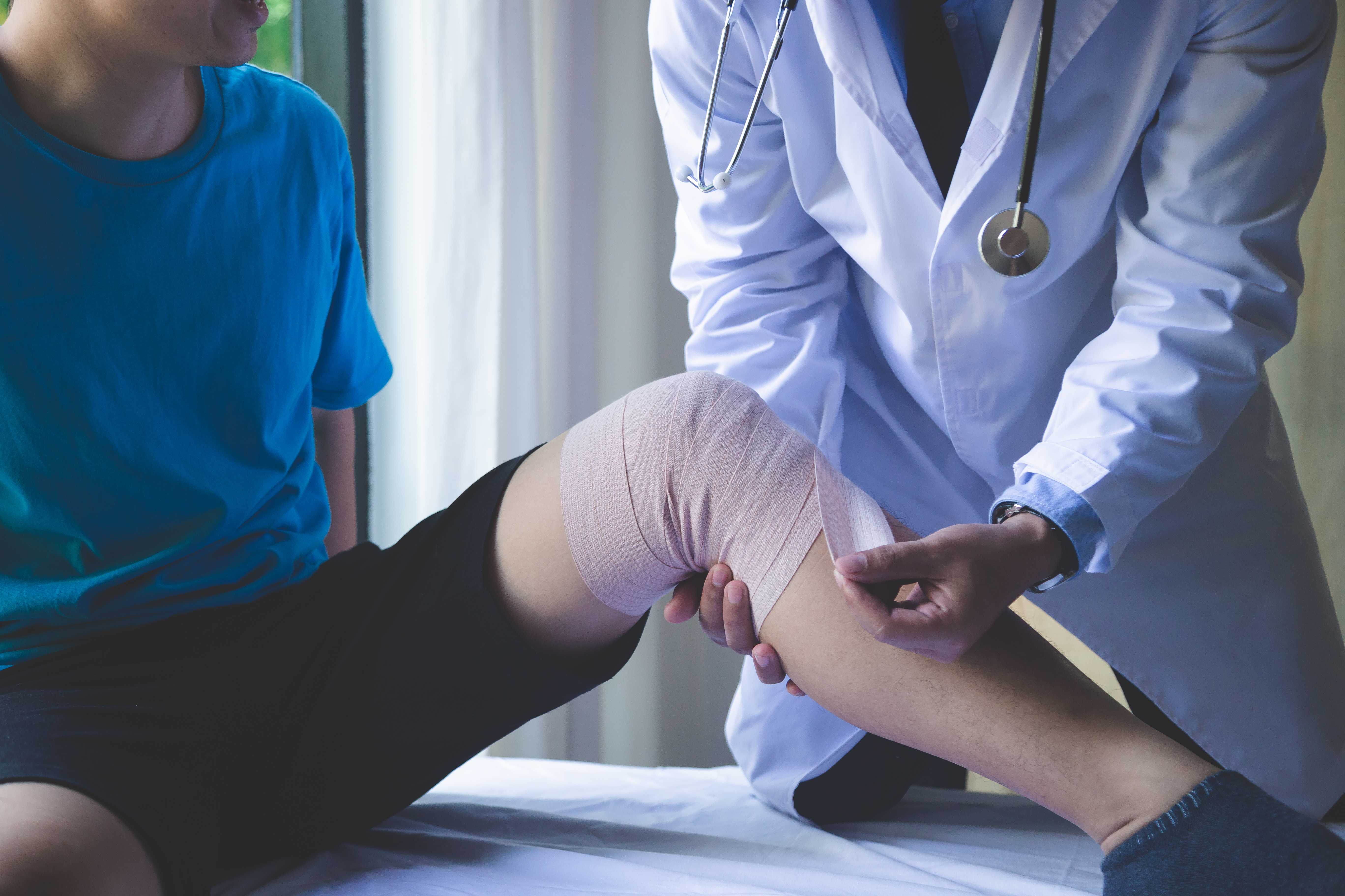
When patients hear "joint surgery," most assume it means replacement, removing the damaged joint and fitting a prosthesis. But there is a substantial and often overlooked middle ground between "keep managing with tablets" and "replace the whole joint."
That middle ground is a joint reconstruction family of procedures that aim to preserve, restore, or realign the natural joint rather than replacing it. For younger patients, active patients, and those with specific patterns of joint disease, reconstruction procedures can provide excellent pain relief, restore function, and delay or sometimes eliminate the need for joint replacement by years or even decades.
This guide covers what joint reconstruction means in practice, which procedures fall under this umbrella, who is a suitable candidate, and how reconstruction fits into the overall management pathway for joint disease.
What Is Joint Reconstruction?
Joint reconstruction refers to surgical procedures that correct, restore, or optimise the natural joint without replacing it with a prosthesis. It differs from joint replacement in a fundamental way: the goal is to preserve and improve the existing joint structure, not to replace it. The primary rationale for joint reconstruction over replacement:
Implant limitations in young patients: Modern joint replacements are excellent, but they are not designed to last indefinitely in very active patients. A 35-year-old who undergoes total knee replacement will almost certainly need a revision (redo) surgery in their 50s or 60s, and revision surgery is more complex than primary replacement. Every year that a reconstruction procedure delays replacement is a year of better function before that revision becomes necessary.
Preservation of bone stock: Joint replacement requires the removal of significant bone. Reconstruction procedures preserve bone, making any future replacement simpler and more likely to succeed.
More natural joint feel: Many patients who have joint replacement describe the knee or hip as "functional but not the same." A reconstructed natural joint, in patients where the reconstruction succeeds, typically feels more natural.
Activity level: Joint replacement restricts certain high-impact activities, such as running, jumping, and contact sports, to protect the implant. Reconstruction procedures typically allow a return to higher activity levels.
High Tibial Osteotomy (HTO) - The Most Important Joint Reconstruction Procedure
High tibial osteotomy is the most commonly performed and most evidence-supported joint reconstruction procedure for the knee.
1. What HTO Is
HTO is a surgical procedure that cuts and realigns the tibia (shin bone), changing the mechanical axis of the leg to shift load away from the arthritic compartment of the knee.
Here's the principle: in most patients with medial compartment knee osteoarthritis, the leg is slightly bow-legged (varus alignment). This means that when standing and walking, the body's weight passes through the inner (medial) side of the knee, which happens to be the arthritic side. Every step loads the damaged compartment further, accelerating the arthritis.
HTO cuts the tibia just below the joint and creates a wedge, either removing a wedge (closing wedge) or opening a wedge on the inner side and filling it with bone graft, to correct the leg alignment from varus to a neutral or slightly valgus (mild knock-knee) position.
This alignment correction shifts the weight-bearing axis to the outer (lateral) side of the knee, the side with healthy cartilage. The arthritic inner compartment is "unloaded." Pain decreases because the damaged cartilage is no longer being compressed with every step.
2. Who Is HTO For?
HTO is specifically appropriate for:
- Younger patients (typically under 60) with isolated medial compartment knee OA and varus alignment.
- Patients with good overall joint health, except for the medial compartment, the lateral compartment, and the cartilage elsewhere, must be in reasonable condition.
- Active patients who want to continue working, playing sports, and living an active life, and who are not candidates for replacement yet.
- Patients who have had a previous meniscal tear or meniscectomy and have developed post-meniscectomy arthritis.
3. What Happens During HTO
Under general or spinal anaesthesia, an incision is made below the knee on the inner side. The tibia is carefully cut, partially through the bone, in the opening wedge technique. The cut is gradually opened to the planned degree of correction (typically 5–10 degrees), confirmed with X-ray guidance. A special locking plate and screws hold the correction while the bone heals.
Surgery duration: 60–90 minutes.
Hospital stay: 2–3 days.
4. Recovery After HTO
- Week 1–6: The leg is protected. Walking with a frame, partial weight-bearing as directed by the surgeon.
- Week 6–12: Progressive weight-bearing and physiotherapy.
- Month 3–6: Return to most daily activities and light sports.
- Month 6–12: Return to higher-demand sport for appropriate patients.
- Full bone healing of the osteotomy: 3–4 months typically.
5. How Long Does HTO Last?
Studies show that well-performed HTO provides significant pain relief for 8–12 years in well-selected patients. After this, some patients develop further arthritis and require knee replacement, but at that point, they are older, the replacement will last longer, and the surgery is technically easier in a preserved knee. A patient who has HTO at 45 and knee replacement at 57 has had 12 years of active, unrestricted life between the two procedures, which is a meaningful outcome.
Cartilage Repair Procedures
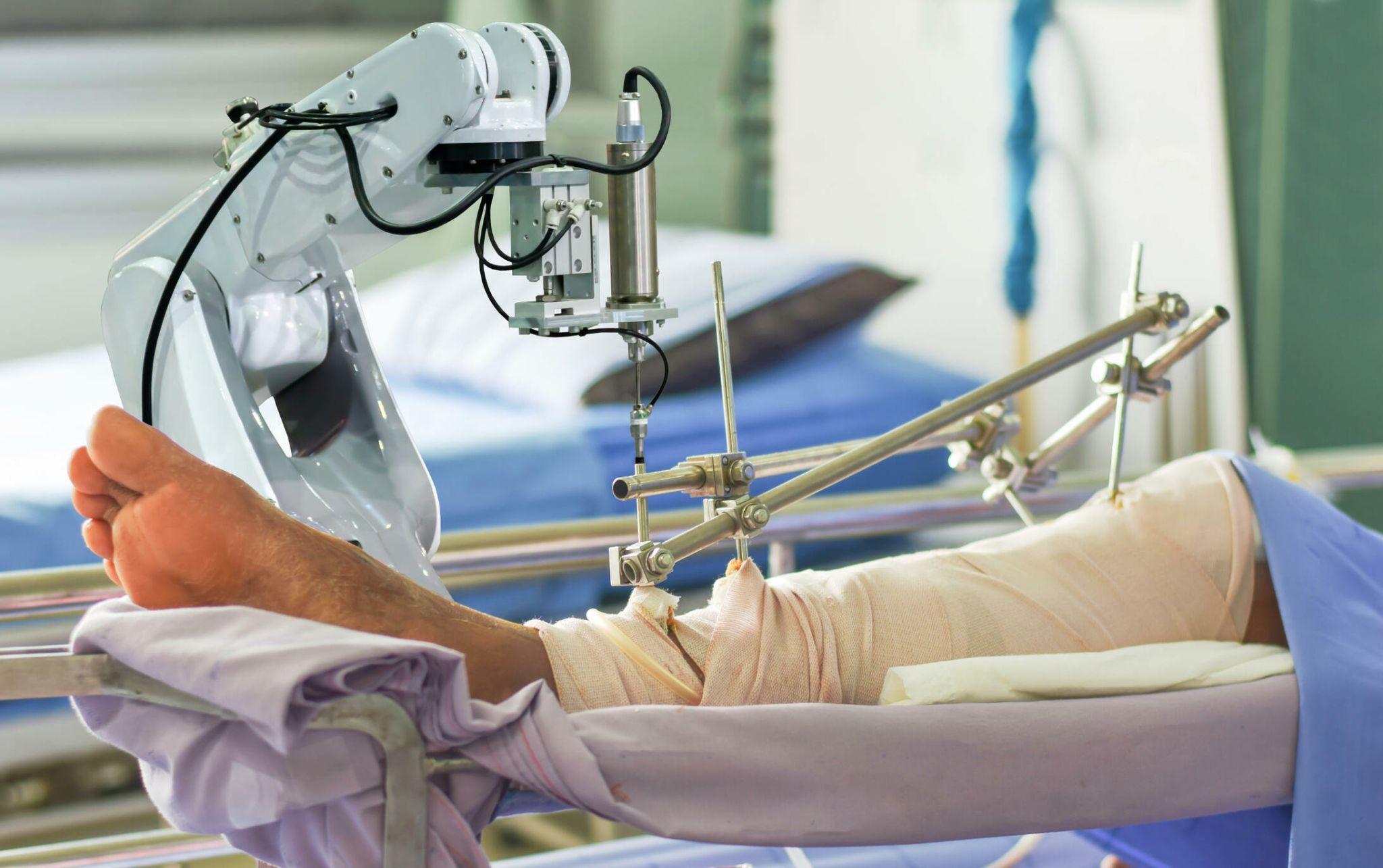
Robotic knee replacement surgery using advanced robotic arm technology. Precision-guided knee joint replacement for improved alignment and recovery
Articular cartilage, the smooth surface covering the ends of bones in a joint, has very poor healing capacity. It has no blood supply of its own, receives nutrition only through diffusion from joint fluid, and has essentially no capacity for regeneration after significant damage. When cartilage defects occur from trauma, from osteochondritis dissecans (OCD), or in early-stage localised arthritis, several procedures can stimulate repair or transplant healthy cartilage to restore the joint surface.
1. Microfracture
The simplest and oldest cartilage repair technique. Multiple small holes are made in the bone beneath the cartilage defect using a small pick. This disrupts the bone marrow, releasing stem cells and growth factors into the defect area. A clot forms, and over months, this clot is gradually replaced by fibrocartilage, a type of cartilage that is not identical to native articular cartilage (which is hyaline cartilage) but provides a degree of joint surface coverage.
Best for: Small, contained cartilage defects (< 2 cm²) in younger patients. Poor results in large defects and in older patients.
Limitations: Fibrocartilage is inferior to hyaline cartilage; it wears faster and provides less durable coverage. Results may deteriorate after 3–5 years in some patients.
2. OATS (Osteochondral Autograft Transfer System)
Plugs of healthy cartilage and bone are harvested from a lower-load area of the patient's own knee (typically the inner margin of the femoral condyle where contact forces are lower) and transplanted into the cartilage defect.
Advantage: Uses the patient's own hyaline cartilage — structurally superior to fibrocartilage. Good results for medium-sized defects.
Limitation: Limited tissue available (donor site morbidity). Best for defects up to 4 cm².
3. Osteochondral Allograft Transplantation
Similar to OATS, but the cartilage and bone plugs come from a cadaveric donor rather than the patient's own knee. This allows treatment of larger defects without the donor site limitation.
Advantage: No limit on defect size. Fresh allograft (used within days of harvesting) has the best chondrocyte viability.
Limitation: Availability of fresh allografts. Risk of immune reaction and disease transmission (very low but present).
4. MACI (Matrix-Induced Autologous Chondrocyte Implantation)
A two-stage procedure:
- First surgery: Small cartilage biopsy taken from a low-load area of the knee and sent to a laboratory. Over 4–6 weeks, the chondrocytes (cartilage cells) are grown in culture on a special membrane (matrix).
- Second surgery: The cell-seeded matrix is implanted into the cartilage defect and fixed in place.
Advantage: Uses the patient's own cartilage cells. Better tissue quality than microfracture. Good for medium-to-large defects (2–10 cm²). Best outcomes in younger patients.
Limitation: Two surgeries required. Currently limited availability in India. Expensive. Long rehabilitation (12–18 months to full activity).
When Is Joint Reconstruction The Right Choice?
Reconstruction is appropriate when:
- The patient is too young or too active for joint replacement to be the optimal solution.
- The pattern of joint disease is localised, one compartment or a specific cartilage defect, rather than pan-articular (all of the joint) damage.
- There is malalignment contributing to uneven joint loading, which can be corrected with osteotomy.
- Conservative treatment has failed, but the joint is not yet at end-stage degeneration requiring replacement.
- The patient understands and accepts the activity restrictions during recovery and the potential need for further surgery in the future.
Reconstruction is not appropriate when:
- Arthritis is Grade 4 (bone-on-bone) across multiple compartments. Replacement is needed.
- The patient is elderly with limited activity demands, replacement provides excellent outcomes and faster rehabilitation.
- The patient's anatomy, bone quality, or ligament status does not support the planned reconstruction.
Joint Reconstruction Vs. Joint Replacement - How To Decide
This is the conversation that happens at a consultation, and it requires an honest assessment of:
| Factor | Favours Reconstruction | Favours Replacement |
|---|---|---|
| Age | Under 55–60 | Over 65 |
| Activity level | High (sport, physical work) | Moderate to low |
| Pattern of arthritis | Isolated compartment, localised | Multi-compartment, pan-articular |
| Bone quality | Good | Osteoporotic (replacement cement fixation preferable) |
| Alignment deformity | Significant varus (favours HTO) | Fixed deformity (replacement corrects it) |
| Patient expectations | Willing for longer recovery, understands it may not be permanent | Wants definitive, predictable solution |
The honest surgeon will have this conversation with every patient rather than defaulting to replacement because it is technically more straightforward.
Joint Reconstruction In Noida - Dr. Mayank Chauhan At Prakash Hospital
Dr. Mayank Chauhan, Senior Orthopedic Surgeon at Prakash Hospital, Sector 33, Noida, evaluates each patient for the full spectrum of options from non-surgical management through joint reconstruction to joint replacement, matching the treatment to the patient's specific anatomy, disease pattern, age, and activity goals.
For younger patients in Noida who have been told they "need a knee replacement" but are in their 40s or early 50s, a second opinion that specifically evaluates whether HTO or another joint reconstruction procedure is appropriate is often worthwhile. To book a consultation, call the number listed on the website.
The Bottom Line

Orthopedic doctor explaining a knee joint model to a patient in clinic
Joint reconstruction surgery, High Tibial Osteotomy, cartilage repair, and related procedures occupy an important and often-overlooked space between conservative management and joint replacement. For the right patient, it provides excellent pain relief, allows a return to active life, and meaningfully delays or avoids the need for replacement.
The key is patient selection, matching the procedure to the specific joint disease, patient age, alignment, and activity demands. Not every patient is suitable. But for those who are, joint reconstruction can be genuinely life-changing.
To consult Dr. Mayank Chauhan, Senior Orthopedic Surgeon in Noida, call the number listed on the website.