Sciatica Vs Slip Disc - Is There A Difference? What You Need To Know

A middle-aged man sits on a couch, twisting slightly as he holds his lower back in pain. A glowing red highlight on his back visually emphasizes the area of discomfort, suggesting sciatica, lumbar strain, or nerve irritation. The room is bright with indoor plants and shelves in the background.
Walk into any orthopedic clinic in Noida, and you'll meet patients who use "sciatica" and "slipped disc" interchangeably. Friends who've been told they have sciatica wonder if they need an operation for their disc. Patients with an MRI showing a herniated disc call it "my sciatica." Family members advise each other based on what "works for sciatica" without knowing whether the underlying cause is the same.
This confusion is not trivial. The distinction between sciatica (a symptom) and a slipped disc (a diagnosis) is clinically important because not all sciatica comes from a disc, and not all slipped discs produce sciatica. Getting treatment right depends on understanding which one you actually have, and what's causing it.
The Core Distinction - Symptom Vs. Diagnosis
Sciatica is a symptom. Slipped disc (herniated disc) is a diagnosis. This is the essential clarification. Sciatica describes a specific type of pain that radiates along the path of the sciatic nerve, from the lower back through the buttock and down the back of the leg, sometimes reaching into the calf and foot. It may be accompanied by numbness, tingling, or weakness in the leg.
A slipped disc (medically: herniated or prolapsed intervertebral disc, PIVD) is a structural problem where the soft inner core of an intervertebral disc has pushed through its outer ring and is pressing on a nerve. This is a diagnosis you see on an MRI report.
The relationship between them: A slipped disc is the most common cause of sciatica approximately 90% of sciatic episodes are caused by lumbar disc herniation. But sciatica can also be caused by several other conditions that have nothing to do with a disc.
Practical implication: When a doctor tells you "you have sciatica," they are describing your symptoms. When they tell you "you have a herniated disc at L4-L5," they are telling you what's causing those symptoms. These are two different pieces of information. Both matter, but they answer different questions.
What Is The Sciatic Nerve?
The sciatic nerve is the largest in the human body. It originates from the lower lumbar spine (from nerve roots L4, L5, S1, S2, S3) and passes through the buttock, down the back of the thigh, dividing behind the knee into the tibial nerve (going into the calf and sole) and the common peroneal nerve (going to the outer lower leg and top of the foot).
When the sciatic nerve or any of its contributing nerve roots is compressed, irritated, or inflamed, pain travels along its path. Depending on which nerve root is involved, the pain follows a slightly different pattern:
- L4 nerve root: Pain down the inner lower leg to the inner foot. Weakness with knee extension.
- L5 nerve root (most common): Pain down the outer thigh, outer calf, to the top of the foot and big toe. Weakness with foot dorsiflexion (lifting the front of the foot).
- S1 nerve root (second most common): Pain down the back of the thigh and calf to the heel and outer foot. Weakness with plantar flexion (pushing down the foot). Absent ankle reflex.
An experienced clinician can often identify which nerve root is involved from the pattern of symptoms, before an MRI confirms it.
Causes Of Sciatica - Beyond The Slipped Disc
While lumbar disc herniation causes the majority of sciatic episodes, it is not the only cause. Several other conditions can produce identical or very similar sciatic symptoms, and each has different treatment implications.
1. Lumbar Disc Herniation (Slip Disc) - Most Common Cause

Medical illustration showing a herniated spinal disc pressing on a nerve, with some diagrams of a human body showing the spine and areas of pain in the back.
The classic mechanism. The herniated disc material presses directly on the nerve root as it exits the spinal canal. Produces the sharp, electric-shock type of leg pain that patients describe as "shooting down the leg." Typically worse with sitting and forward bending, better with lying flat. An MRI confirms the level and degree of herniation.
2. Spinal Stenosis
Narrowing of the spinal canal from degenerative changes, thickened ligaments, bulging discs, and enlarged facet joints can compress nerve roots without a specific disc herniation. More common in patients over 60. Produces neurogenic claudication leg pain that comes on with walking (particularly uphill or on flat ground) and is relieved by sitting or bending forward. This is distinct from disc-related sciatica in an important way: disc sciatica is typically worse with sitting; stenosis sciatica is worse with walking and better with sitting.
3. Piriformis Syndrome
The piriformis is a muscle in the buttocks that the sciatic nerve runs through (in some people) or directly beneath. When the piriformis muscle is tight, in spasm, or irritated from prolonged sitting, hip overuse, or trauma, it can compress the sciatic nerve at this level.
Piriformis syndrome produces buttock pain with radiation down the leg, very similar to disc-related sciatica. Key differentiator: There is no spine pathology on MRI, but there is localised tenderness over the piriformis muscle in the buttock, and specific hip movements provoke symptoms. More common in cyclists, runners, and people who sit for prolonged periods. It is important because treatment is completely different, targeted stretching and physiotherapy for the piriformis muscle, not disc treatment.
4. Spondylolisthesis
When a vertebra slips forward on the one below it, it can narrow the foramina (the exit holes through which nerve roots pass), compressing a specific nerve root. This produces both back pain and leg pain similar to a disc herniation.
5. Sacroiliac (SI) Joint Dysfunction
The sacroiliac joint connects the sacrum (base of the spine) to the pelvis. When inflamed or dysfunctional, it can produce pain that radiates into the buttock and upper posterior thigh, mimicking sciatica. However, SI joint pain does not typically radiate below the knee, which can help differentiate it clinically.
6. Lumbar Facet Joint Arthritis
Inflamed facet joints can refer pain into the buttock and thigh, mimicking early sciatica. Facet joint pain is typically localised to the lower back and buttocks, does not usually radiate below the knee, and is worsened by extension (backward bending) of the spine.
7. Vascular Claudication
Not a spinal cause at all, pain in the legs from peripheral arterial disease (narrowed arteries) can mimic neurogenic claudication. Key difference: Vascular claudication is relieved by stopping walking (standing still); neurogenic claudication requires sitting or bending forward. A vascular assessment (ankle-brachial index) differentiates these.
8. Tumours
Rare, but important to mention. Tumours within the spinal canal (primary or metastatic) can cause progressive leg pain and neurological symptoms. Red flags: bilateral symptoms, pain that is constant and worse at night, unexplained weight loss, and a history of cancer.
How Are Sciatica And Slipped Disc Diagnosed?
Step 1: Clinical History
Key questions your orthopedic surgeon will ask:
- Where exactly does the pain start, and where does it go?
- Does it go below the knee? (More likely to be true nerve root compression if it does)
- Is the pain worse with sitting or walking?
- Did it start after a specific event - a lift, a twist, an injury?
- Are there any neurological symptoms - numbness, tingling, weakness?
- Any bladder or bowel changes? (Red flag - possible cauda equina)
Step 2: Physical Examination
The Straight Leg Raise (SLR) test is the most important clinical test for disc-related sciatica. Lying flat, the leg is raised to 30–70 degrees. If this reproduces the sciatic pain down the leg (not just back pain), it is a positive test, strongly suggesting disc herniation compressing the nerve root. Neurological examination of reflexes, sensation in the dermatome distribution, and muscle strength helps identify which nerve root is compressed and at what level.
Step 3: Imaging
MRI is the gold standard. It shows the disc herniation (if present), its level and degree of nerve compression, spinal canal dimensions (for stenosis assessment), and other causes.
X-ray is done routinely to assess overall alignment and rule out fractures or significant spondylolisthesis.
CT scan is used when an MRI is not available or contraindicated.
Electromyography (EMG) and Nerve Conduction Studies may be ordered when the source of nerve dysfunction needs to be further localised, or to exclude peripheral nerve problems.
Treatment - Why The Cause Matters
Disc herniation causing sciatica: Physiotherapy (McKenzie method), NSAIDs, nerve pain medication (pregabalin/gabapentin), epidural steroid injections if inadequate response, microdiscectomy if conservative management fails over 6–8 weeks or if there is progressive neurological deficit.
Spinal stenosis: Physiotherapy (flexion-based exercises opposite to disc herniation treatment), epidural injections, and surgical laminectomy (decompression) when conservative treatment fails.
Piriformis syndrome: Stretching, physiotherapy targeting the piriformis and hip rotators, and ultrasound-guided injection into the piriformis muscle in refractory cases. No spinal surgery.
SI joint dysfunction: Physiotherapy, SI joint injections. No disc surgery.
Vascular claudication: Vascular surgical referral. No spinal treatment.
This is why diagnosis matters. Treating piriformis syndrome with an epidural injection directed at a disc level is not going to help. Treating a patient with spinal stenosis using the extension exercises appropriate for a disc herniation will make their condition worse. And sending someone with vascular claudication for spine surgery is simply wrong.
When Is Sciatica An Emergency?
Most sciatica, even when severe, is not an emergency. But certain features require immediate medical evaluation:
- Loss of bladder or bowel control (difficulty urinating, urinary retention, or incontinence), this is cauda equina syndrome, a surgical emergency. The large disc herniation compressing the cauda equina nerve bundle requires surgery urgently to prevent permanent neurological damage.
- Rapidly progressive leg weakness, weakness that is getting worse over hours to days.
- Bilateral leg symptoms, both legs involved simultaneously.
If any of these are present, go to an emergency department, not a scheduled clinic appointment.
Sciatica And Slipped Disc Treatment In Noida
Dr. Mayank Chauhan, Senior Orthopedic Surgeon at Prakash Hospital, Sector 33, Noida, evaluates all forms of lower back and leg pain, distinguishing disc herniation from other causes of sciatica through careful clinical assessment and appropriate imaging. For patients in Noida and Greater Noida who have been managing leg pain and back pain without a clear diagnosis, a proper evaluation will clarify exactly what is causing the symptoms and determine the most appropriate treatment.
To book a consultation, call the number listed on the website.
The Bottom Line
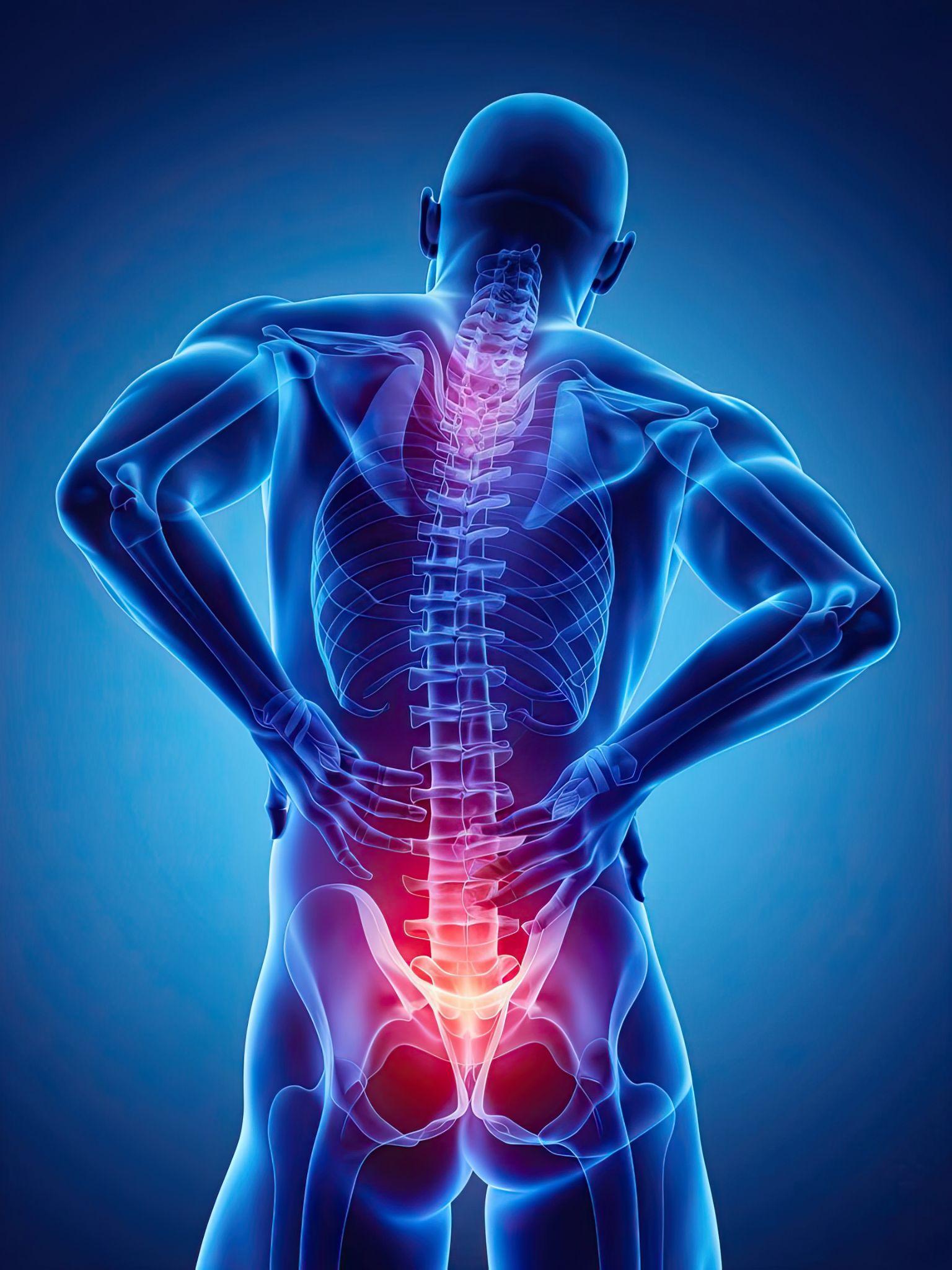
A digital 3D anatomical illustration of a human figure viewed from behind, highlighting the spine. Bright red areas along the lower spine indicate inflammation or nerve compression, commonly seen in sciatica or lumbar pain conditions.
Sciatica is a symptom of radiating leg pain along the sciatic nerve's path. Slip disc is one cause of that symptom, the most common one, but not the only one. Getting the right treatment requires getting the right diagnosis. And getting the right diagnosis requires a proper clinical evaluation, not just an MRI report, and not just a family member's experience with "sciatica."
To consult Dr. Mayank Chauhan, Senior Orthopedic Surgeon in Noida, call the number listed on the website.