Avascular Necrosis (AVN) Of Hip - Causes, Stages, And When Surgery Is Needed

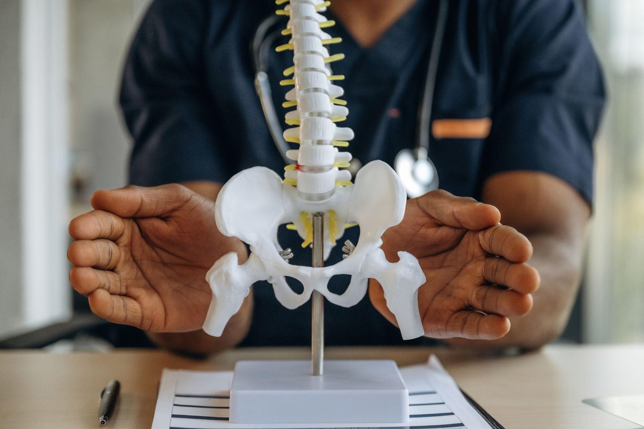
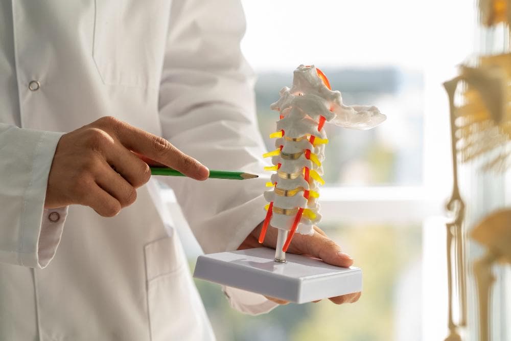
Anatomical model of a hip bone.
Avascular necrosis is a condition that doesn't get talked about enough in India, which is unfortunate, because India has one of the highest rates of AVN in the world. AVN accounts for over 50% of all primary total hip replacement surgeries performed in India, a statistic that would be surprising in most Western countries, where osteoarthritis is the dominant form of arthritis.
More significantly, AVN predominantly affects younger patients; more than 70% of AVN cases in India occur in people between 20 and 40 years of age. It is a disease of young, working-age adults that, if missed or poorly managed, can lead to hip joint destruction within 2 years of onset. Understanding AVN, what causes it, how it progresses, and what treatment at each stage looks like is genuinely important for patients in Noida and Greater Noida who present with unexplained hip or groin pain.
What Is Avascular Necrosis?
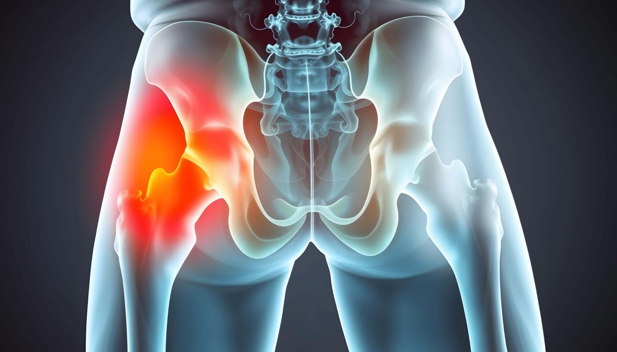
Avascular necrosis (AVN), also called osteonecrosis or ischemic bone necrosis, occurs when the blood supply to a bone is disrupted. Without an adequate blood supply, the bone tissue begins to die. In the hip, this affects the femoral head, the ball of the ball-and-socket joint.
As the bone dies, it loses its structural integrity. The femoral head, which must bear 2.5–3 times body weight with each step, begins to collapse under this mechanical load. Once collapse occurs, the joint surface becomes irregular, articular cartilage is destroyed, and the hip develops secondary arthritis. The timeline from the onset of AVN to femoral head collapse, without intervention, is typically 12–24 months. This is why early diagnosis and prompt treatment are critical.
Causes Of AVN - Why It Is So Prevalent In India
AVN can result from any condition that disrupts blood flow to the femoral head. In India, the most common causes are distinct from those seen in Western countries.
1. Corticosteroid Use (Most Common Cause In India)
Long-term or high-dose corticosteroid use is the single most common cause of AVN in India. Corticosteroids are used extensively in India for:
- Nephrotic syndrome (a kidney disease very common in the Indian population)
- Rheumatoid arthritis and other autoimmune conditions
- COVID-19 treatment (a significant wave of steroid-related AVN followed India's COVID-19 pandemic, with many patients developing bilateral AVN after high-dose steroid protocols)
- Asthma
- Inflammatory bowel disease
- Various other conditions managed with prednisolone or dexamethasone
The mechanism: Corticosteroids cause fat cells to enlarge within the bone marrow, compressing the small blood vessels that supply the femoral head. They also promote fat emboli that block the vessels. The risk is dose-dependent and time-dependent, but AVN can develop even after relatively short high-dose courses.
2. Alcohol Use
Chronic heavy alcohol consumption is the second most common cause. Similar to steroids, alcohol causes fatty infiltration of bone marrow and increases the risk of fat emboli. Alcoholic AVN is almost universally bilateral (both hips).
3. Trauma
Fractures of the femoral neck, the slender part of the femur between the ball and the shaft, can disrupt the blood vessels that run along the neck to the femoral head. Post-traumatic AVN can develop months to years after the original fracture. Hip dislocations also disrupt the blood supply. AVN after hip dislocation develops in a significant proportion of patients, and the risk increases with the delay to reduction (putting the joint back in).
4. Idiopathic (No Identifiable Cause)
In some patients, no specific cause is identified despite thorough investigation. These idiopathic cases account for approximately 20% of AVN.
5. Other Causes
- Sickle cell disease - sickling of red blood cells blocks small vessels
- Coagulation disorders - hypercoagulable states increase thrombotic risk in the femoral head vessels
- Radiation therapy - radiation to the hip area (for pelvic tumours) damages blood vessels
- Decompression sickness - in deep-sea divers (less relevant in Delhi NCR)
- Gaucher's disease - a metabolic storage disorder
The Four Stages Of AVN - The Ficat Classification
AVN progresses through distinct stages, and the appropriate treatment depends critically on the stage at which the patient presents.
1. Stage I - Pre-collapse, Normal X-ray
The femoral head is completely normal on plain X-ray. However, changes are occurring at the cellular level, such as bone marrow oedema and early bone death. MRI detects these changes before any structural abnormality is visible.
Symptoms: Often none or mild hip pain. Many patients at this stage are asymptomatic.
The critical window: Stage I is the optimal time for joint-preserving intervention, the blood supply can potentially be restored before irreversible structural damage occurs.
2. Stage II - Pre-collapse, Abnormal X-ray
X-ray now shows changes in sclerosis (increased bone density), cystic changes, or mixed patterns in the femoral head. But the shape of the femoral head is still intact, no collapse yet. MRI shows the full extent of the necrotic zone.
Symptoms: Hip pain typically in the groin, sometimes radiating into the thigh. Pain with weight-bearing and activity.
Stage III - Collapse
The necrotic bone has lost its structural integrity, and the subchondral bone (the thin layer of bone just beneath the cartilage) fractures, producing the classic "crescent sign" on X-ray. The femoral head begins to flatten and collapse.
Symptoms: Significant hip pain, increasingly constant, limiting walking distance.
Critical point: Once collapse has occurred, the opportunity for joint-preserving surgery is limited. The collapsed head cannot be restored to a perfectly spherical shape.
Stage IV - Arthritic Change
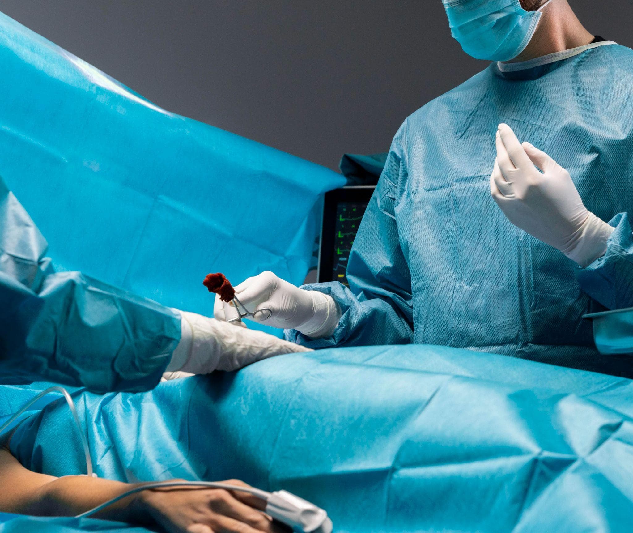
Orthopedic surgeons performing a hip replacement procedure.
Complete collapse of the femoral head with secondary arthritis, narrowing of the joint space, damage to the acetabular cartilage (the socket side), and osteophyte formation. This is end-stage hip disease requiring joint replacement.
Diagnosis
1. Clinical Presentation
AVN should be suspected in any patient, particularly a young adult, presenting with hip or groin pain, especially if they have any of the risk factors described above (steroid use, alcohol use, previous fracture or dislocation).
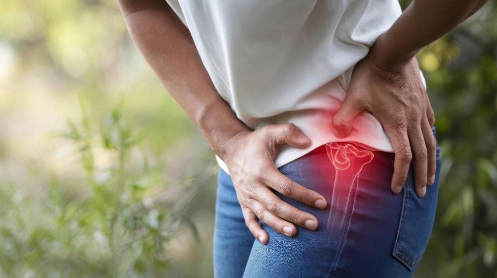
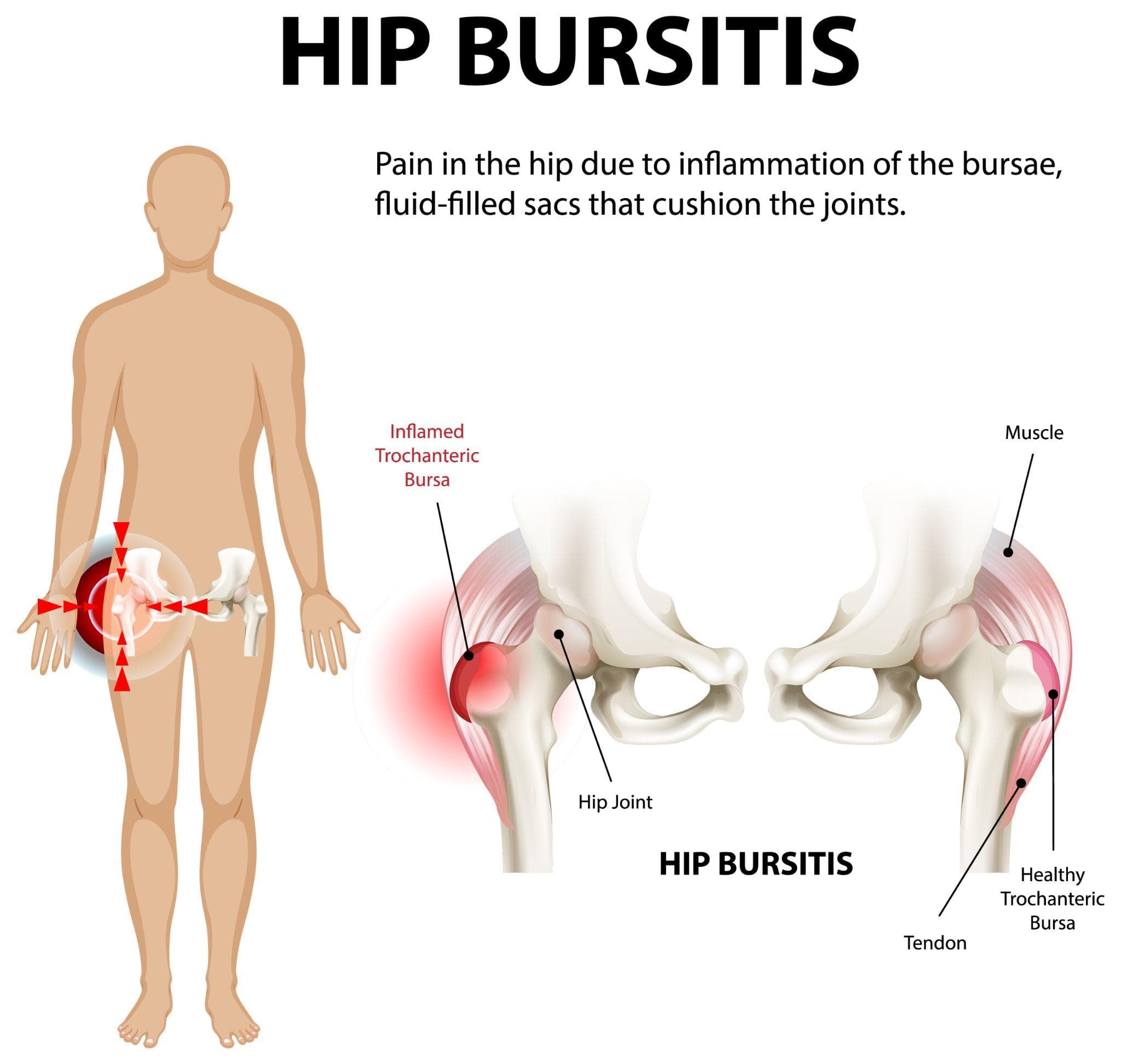
The classic symptom: Groin pain (not lateral hip pain that is usually trochanteric bursitis or IT band pathology). Pain may radiate to the anterior thigh and sometimes the knee. Pain with internal rotation of the hip is a characteristic early finding on examination.
2. Investigations
X-ray: Adequate for Stage II–IV. Stage I is invisible on plain X-ray.
MRI: The gold standard for early AVN (Stage I). Shows the characteristic "double line sign," a specific MRI appearance of the necrotic zone. Also quantifies the extent of involvement (percentage of the femoral head affected), critical for prognosis and surgical planning.
CT scan: Useful for assessing the subchondral bone detail, particularly for detecting the crescent sign (subchondral collapse) and for pre-surgical planning.
Bone scan: Can detect bilateral AVN when only one side is symptomatic. This is important because bilateral AVN is very common (particularly in steroid and alcohol-related AVN), and the contralateral hip should always be assessed.
Treatment - Stage-Based Approach
1. Stage I And Early Stage II - Joint Preservation
Core Decompression: The most widely used joint-preserving procedure. Small channels are drilled into the femoral head through the femoral neck under X-ray guidance. This:
- Relieves elevated intraosseous pressure (which contributes to further vascular compromise)
- Creates channels through which new blood vessels can grow (angiogenesis)
- Allows the introduction of bone graft or biological agents
Done as a minimally invasive procedure (small incision, 1–2 days hospital stay). Most effective for small lesions in Stage I and early Stage II, with success rates of 70–85% in preventing progression in well-selected patients.
Core Decompression with Bone Grafting: Bone graft (from the patient's own pelvis or from a donor) is packed into the channels created during core decompression. The graft provides structural support and stimulates biological repair. More effective than core decompression alone for larger lesions.
Vascularised Fibula Graft: A more complex procedure where a segment of fibula (with its blood supply intact) is transferred to the femoral head. The vascular pedicle restores direct blood supply to the necrotic zone. Best results in young patients with Stage II AVN and medium-sized lesions. Technically demanding and requires a specialised surgical team.
Bisphosphonate Therapy: Bisphosphonates (alendronate, zoledronic acid) reduce bone resorption and may slow the progression of AVN, particularly in early stages. The evidence is moderate. Often used as an adjunct to surgical procedures rather than as a standalone treatment.
Stem Cell Therapy: Autologous bone marrow-derived stem cells (harvested from the patient's own iliac crest) are injected into the necrotic zone, often combined with core decompression. The stem cells potentially stimulate angiogenesis and bone repair. Evidence is growing; several Indian studies have shown promising results. It is not yet a fully established standard of care, but it is increasingly offered at specialised Indian orthopaedic centres.
Late Stage II And Stage III - Limited Options
Once the femoral head shows early collapse (Stage III), joint-preserving options are technically possible but have more limited predictability. Osteotomy procedures (realigning the hip to shift load away from the necrotic segment) can be performed in selected younger patients at Stage II–III, buying time before replacement becomes necessary.
The decision at this stage requires a detailed conversation between the surgeon and patient about realistic expectations, age, activity demands, and the patient's acceptance of further surgery in the future.
Stage IV - Total Hip Replacement
End-stage AVN with collapsed head and established arthritis requires total hip replacement, the definitive treatment for pain relief and function restoration. For AVN patients, several considerations apply:
- AVN patients are often young; the hip replacement must be planned with the assumption that revision surgery may be needed in 15–25 years.
- Implant selection should favour long-lasting bearing surfaces (ceramic-on-ceramic or ceramic-on-polyethylene).
- Uncemented fixation is preferred in younger patients with good bone quality.
- Bilateral AVN (very common) may require staged bilateral hip replacement.
The outcomes of total hip replacement for AVN are consistently excellent, comparable to or better than replacement for OA, with high patient satisfaction and dramatic pain relief.
Bilateral AVN - An Important Point
Because AVN is frequently bilateral, particularly in steroid-related and alcohol-related cases, both hips must be evaluated at initial presentation. A patient with known AVN in one hip who has not had the other hip imaged has an incomplete assessment. Bilateral MRI is appropriate in all non-traumatic AVN patients.
AVN Treatment In Noida - Dr. Mayank Chauhan At Prakash Hospital
Dr. Mayank Chauhan, Senior Orthopedic Surgeon at Prakash Hospital, Sector 33, Noida, manages AVN of the hip across all stages from early core decompression procedures for Stage I–II disease through to total hip replacement for end-stage AVN.
For patients in Noida and Greater Noida presenting with unexplained groin pain, particularly those with a history of steroid use, alcohol use, or previous hip trauma, early MRI evaluation is essential. Early diagnosis at Stage I or II offers the best chance of joint preservation. To book a consultation, call the number listed on the website.
The Bottom Line
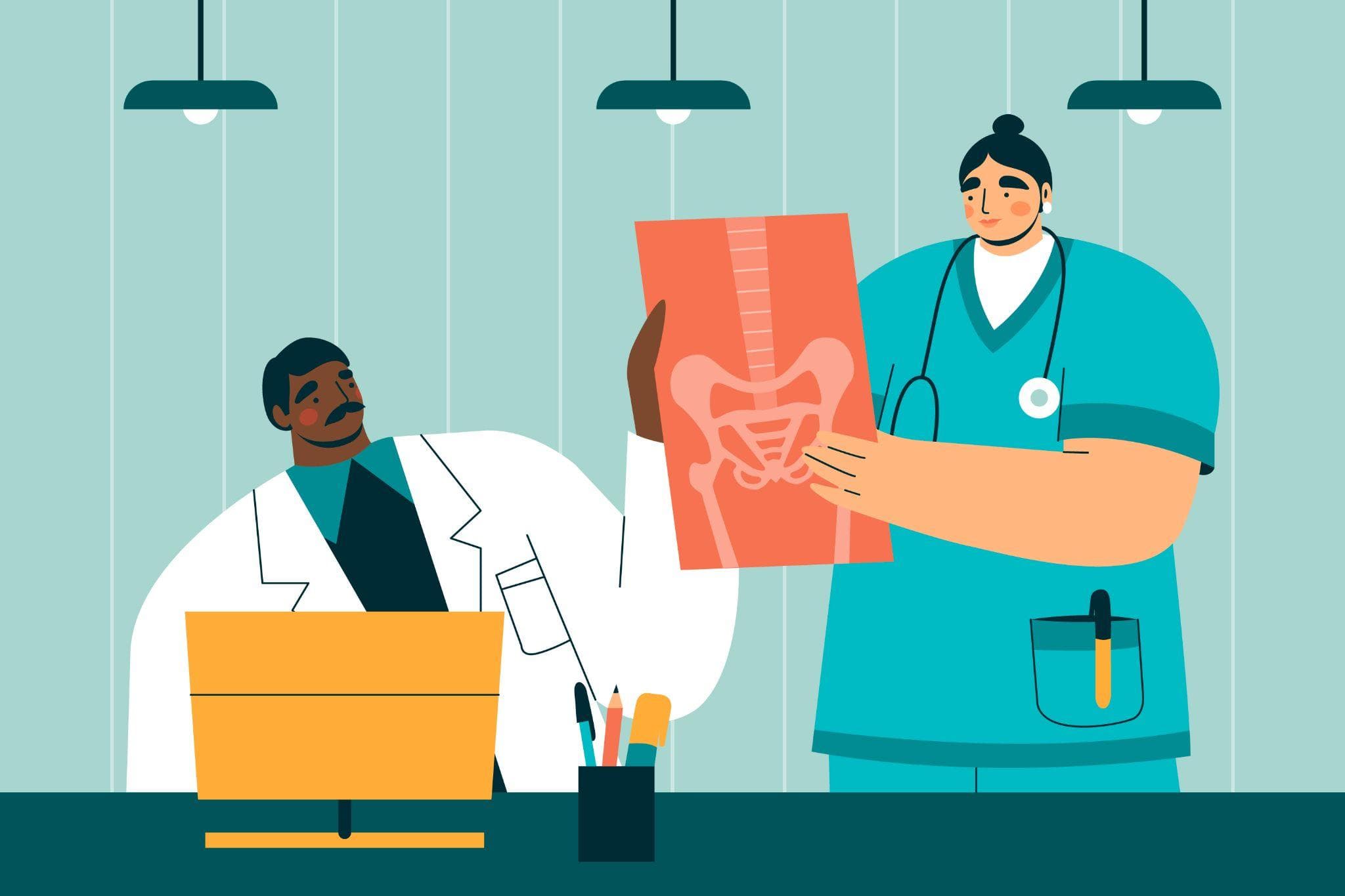
An illustration showing a doctor and a nurse holding a hip X-ray together in a clinic.
Avascular necrosis of the hip is common in India, predominantly affects young working-age adults, and progresses rapidly if untreated. Early diagnosis when MRI shows Stage I disease offers the best chance of joint-preserving intervention. Late diagnosis when the femoral head has collapsed inevitably leads to hip replacement at a young age.
If you have unexplained groin or hip pain, particularly with risk factors like steroid use, alcohol use, or previous hip injury, get an MRI. The window for preservation is narrow. To consult Dr. Mayank Chauhan, Senior Orthopedic Surgeon in Noida, call the number listed on the website.