PCL Tear - Causes, Symptoms, Diagnosis, And Treatment

A diagram is shown, illustrating PCL injury on the knee.
When people talk about knee ligament injuries, the ACL takes almost all of the attention. With good reason, it is the most commonly surgically treated ligament injury in sport. But the PCL, the posterior cruciate ligament, is injured more often than most people realise, and is consistently underdiagnosed in emergency departments and primary care settings across Noida and Delhi NCR.
A 2025 expert consensus from 17 countries on PCL injury management highlights a fundamental challenge: despite being a relatively common musculoskeletal condition, there is currently a lack of consensus on decision-making and treatment for PCL injuries. This makes accurate diagnosis and proper specialist assessment even more important because the management pathway for PCL injuries is meaningfully different from ACL injuries, and getting it wrong leads to chronic knee problems. This blog explains what the PCL is, how it is injured, what the clinical picture looks like, and how it is managed from mild sprains to complete ruptures.
The PCL - The Strongest Ligament In The Knee
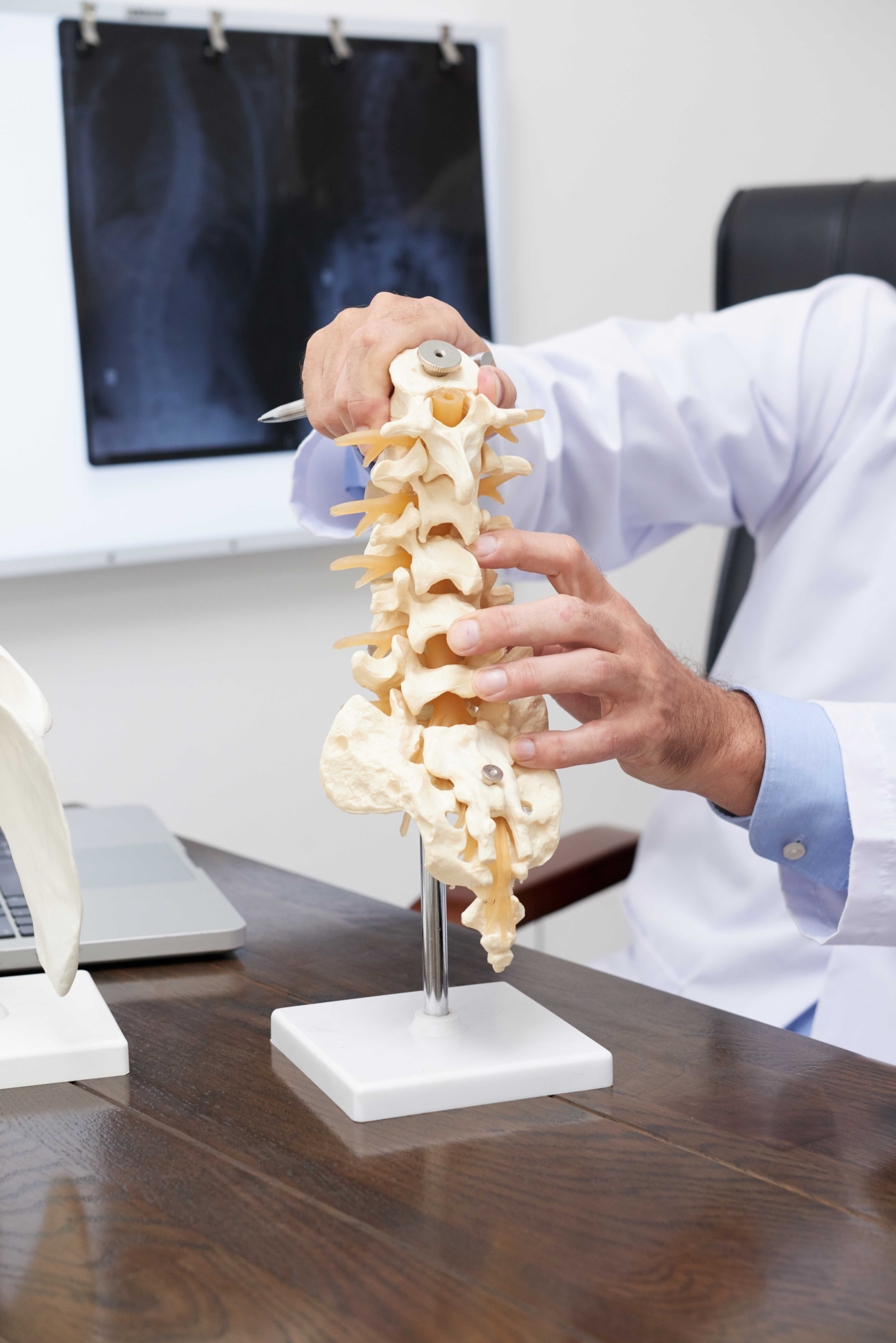
The posterior cruciate ligament (PCL) runs through the centre of the knee joint diagonally, in the opposite direction to the ACL. The two cruciate ligaments cross each other, which is the origin of the term "cruciate" (from the Latin for cross). What the PCL does:
- Prevents the tibia (shin bone) from sliding backward on the femur (thigh bone), posterior tibial translation.
- Provides rotational stability.
- Works in combination with the ACL to give the knee its central pivot stability.
The PCL is significantly larger and stronger than the ACL, approximately twice its tensile strength. This is why PCL injuries are less common than ACL injuries; it takes substantially more force to rupture the PCL.
When the PCL is most stressed: During knee flexion (bending) with posterior-directed force on the tibia, the classic "dashboard mechanism." Also, during hyperflexion (extreme knee bending) and hyperextension injuries.
How PCL Tears Happen
1. The Dashboard Mechanism (Most Classic)
The classic PCL injury mechanism is so named because it was classically described in road traffic accidents, and remains very relevant in Noida's traffic context. When a vehicle occupant hits their bent knee against the dashboard in a collision, the force is directed backward (posteriorly) on the tibia. This posterior force, applied with the knee flexed, directly stresses the PCL. The impact drives the tibia backward, stretching and potentially rupturing the PCL.
In Noida's high-traffic environment, two-wheelers, auto-rickshaws, and cars cause dashboard-type injuries in road accidents, which are a significant cause of PCL tears.
2. Direct Blow To The Anterior Tibia
Any direct blow to the front of the shin with the knee bent, a football tackle, a cricket collision, or a fall that impacts the front of the knee can cause a PCL injury through the same posterior-directed force mechanism.
3. Hyperflexion
Extreme forced knee bending, landing on a bent knee, or a fall forward onto a flexed knee can tear the PCL through the stretching force applied to the posterior structures.
4. Hyperextension
Severe hyperextension (forcing the knee into extreme straightening) can injure both cruciate ligaments. The PCL may be injured in combination with the ACL and posterolateral corner structures.
5. Multi-Ligament Injuries
PCL tears frequently occur alongside other ligamentous injuries. High-energy traumas, such as road accidents and sporting collisions, produce multi-ligament knee injuries where the PCL, ACL, MCL, LCL, and posterolateral corner structures are injured in combination. These complex injuries require specialist assessment and carefully planned management.
Grading PCL Injuries
PCL injuries are graded based on the degree of posterior tibial translation:
Grade 1 (Mild Sprain): Posterior tibial translation of up to 5 mm. Ligament fibres stretched but intact. Knee feels stable.
Grade 2 (Partial Tear): 5–10 mm of posterior translation. The tibial plateau remains in front of the femoral condyles (it doesn't sag behind them). This is an incomplete tear; the ligament has significant structural integrity remaining.
Grade 3 (Complete Tear): More than 10 mm of posterior translation. The tibia sags behind the femur (posterior sag sign). Complete disruption of the ligament.
The critical difference from ACL grading: Even complete (Grade 3) isolated PCL tears are often managed conservatively, unlike ACL tears, which in active patients almost always require reconstruction. This is because the PCL has a better blood supply and healing capacity than the ACL, and because many patients with isolated PCL deficiency can function adequately with proper rehabilitation.
Symptoms Of PCL Injury
PCL injuries are often less dramatic at the moment of injury than ACL tears, which may contribute to their underdiagnosis. Acute phase (immediately after injury):
- Pain in the back of the knee.
- Swelling is typically less rapid than ACL haemarthrosis; it may be moderate rather than dramatic.
- Ability to walk is often preserved (unlike severe ACL tears), which causes many patients to dismiss the injury.
- Stiffness with knee flexion.
Subacute and chronic phase:
- Posterior knee pain, particularly with activities that flex the knee (going down stairs, squatting, cycling).
- A sense that the knee is unstable or "giving way," though this is often less pronounced than ACL deficiency.
- Difficulty with activities requiring knee control at different angles.
- In chronic PCL deficiency, progressive anterior knee pain (patellofemoral pain) can develop as the altered mechanics of the PCL-deficient knee change the loading pattern on the kneecap.
The key clinical finding: The posterior sag sign with the patient supine and both knees bent at 90 degrees, the tibia of the PCL-injured knee "sags" visibly backward compared to the other side. This is pathognomonic of PCL deficiency.
The posterior drawer test: With the knee at 90 degrees of flexion, the tibia is pushed backward. Increased posterior translation compared to the other knee confirms PCL laxity.
Diagnosis
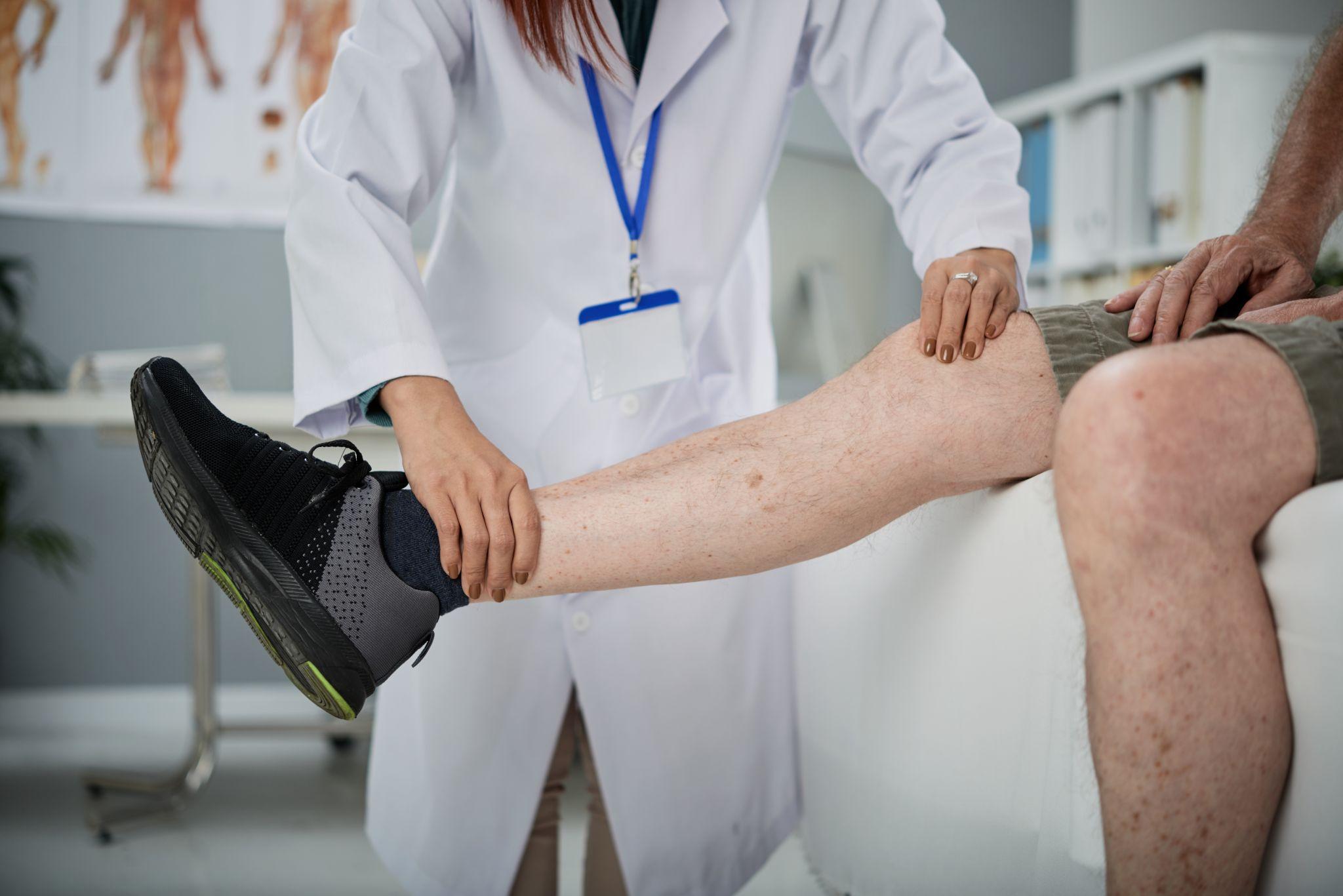
An orthopedic knee doctor examining the knee of a patient.
1. Clinical Examination
An experienced orthopedic surgeon can diagnose a PCL injury clinically in most cases. The posterior sag sign and posterior drawer test are the most reliable and specific tests. The examination must also assess for associated injuries:
- Posterolateral corner (PLC) assessment: palpation of the LCL, dial test (external rotation asymmetry), and varus stress test.
- ACL assessment (often injured alongside)
- MCL assessment (valgus stress)
PCL injuries associated with posterolateral corner injury are particularly important to identify because the PLC must be addressed surgically when the PCL is reconstructed, and failure to identify PLC injury leads to PCL reconstruction failure.
2. MRI
Essential for:
- Confirming the PCL tear and grading its extent
- Identifying associated injuries: posterolateral corner, ACL, MCL, menisci, cartilage
- Surgical planning
MRI sensitivity for PCL tears is approximately 95%; it is the gold standard investigation.
3. Stress X-rays
Weight-bearing and stress radiographs can quantify the degree of posterior tibial translation and help determine the functional severity of the injury, particularly useful in planning surgical reconstruction.
Treatment
1. Conservative Management - The First Line For Most PCL Injuries
Unlike ACL tears, many PCL injuries, including some Grade 3 complete tears, are managed conservatively with good outcomes, particularly in isolated injuries without associated ligamentous damage. The rationale for conservative management: The PCL has a better intrinsic healing capacity than the ACL, related to its better blood supply and the mechanical environment in which it heals. Multiple studies have shown that isolated Grade 1 and Grade 2 PCL tears consistently heal well with rehabilitation. Even some Grade 3 isolated tears in relatively sedentary patients can be managed without surgery.
A 2025 Delphi-based expert consensus from the German Knee Society, involving 90 experienced clinicians from orthopaedics and sports medicine worldwide, confirms that conservative treatment is the first-line approach for isolated PCL injuries, with surgery reserved for specific indications.
Conservative treatment protocol:
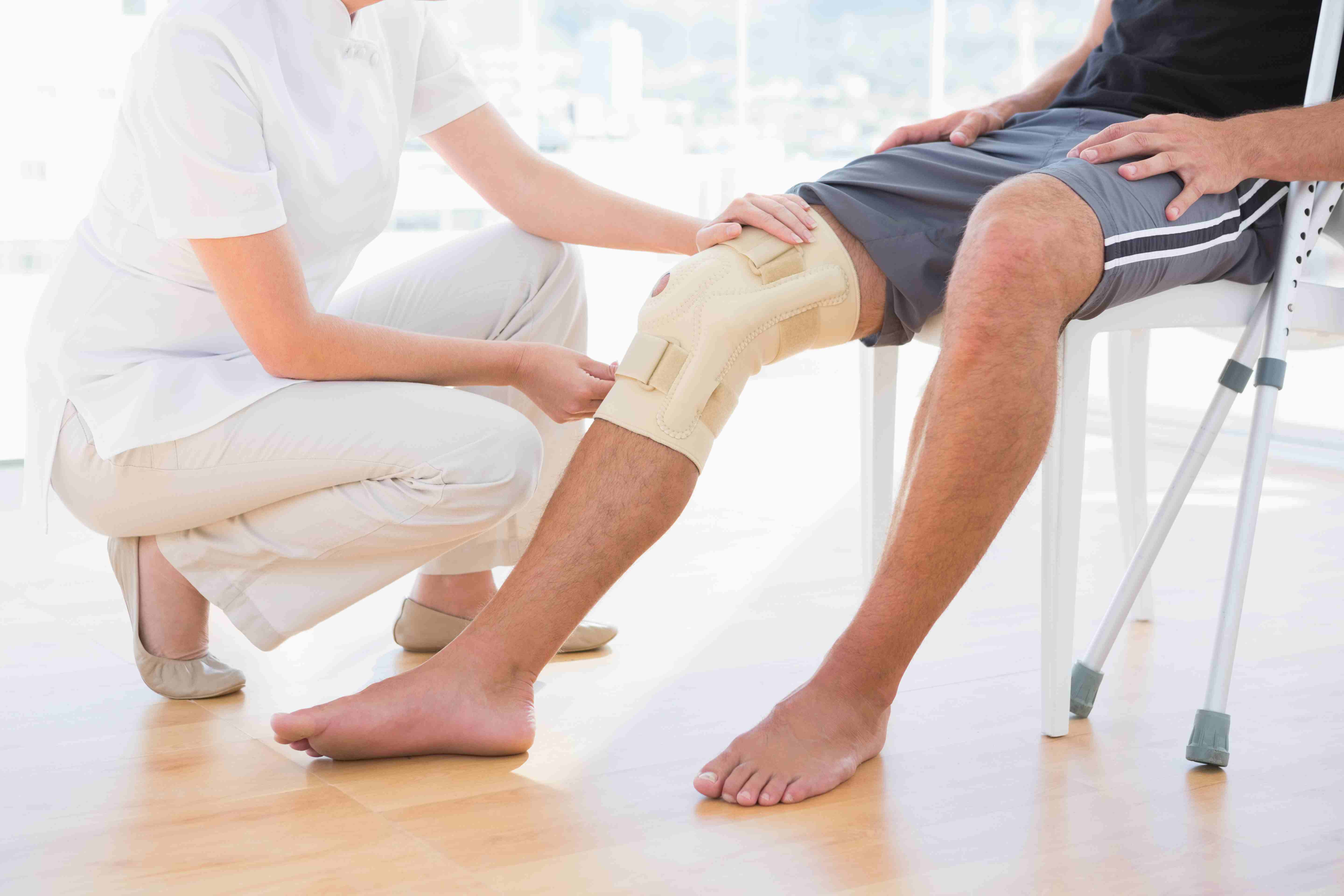
Phase 1 - Acute (Weeks 1–3): A posterior support brace (which prevents the tibia from sagging backward) is worn. This unloads the healing PCL and reduces the posterior tibial sag that would place the torn ligament ends in a poor healing position.
Phase 2 - Quadriceps Rehabilitation (Weeks 3–12): Strong quadriceps muscles are the key dynamic stabiliser of the PCL-deficient knee; they actively pull the tibia forward, compensating for the absent passive restraint of the PCL. Aggressive quadriceps strengthening is the cornerstone of PCL rehabilitation.
Key exercises:
- Closed-chain quadriceps exercises (leg press, step-ups, mini-squats)
- Open-chain quad sets with hamstring co-contraction avoided (hamstring contraction pulls the tibia backward in the same direction as PCL deficiency)
- Cycling is excellent for the quadriceps without hamstring-driven posterior tibial shear
- VMO (vastus medialis oblique) strengthening for patellar tracking
Phase 3 - Return To Sport (Months 3–6): Progressive return to sport-specific activities when the quadriceps symmetry index exceeds 85%, proprioception is restored, and the knee is functionally stable.
Surgical Management - When It Is Indicated
Surgery is reserved for specific situations where conservative management is unlikely to succeed or has failed:
Absolute surgical indications:
- Acute PCL avulsion fractures (where the ligament pulls off a bone fragment), the fragment can often be fixed back directly, with excellent results. This is a bone-to-bone healing situation that is different from mid-substance ligament tears.
- Grade 3 PCL tears associated with posterolateral corner injury. PLC injuries do not heal conservatively; the combined injury requires surgical stabilisation.
- Multi-ligament knee injuries where PCL reconstruction is performed as part of a comprehensive reconstruction.
Relative surgical indications:
- Grade 3 isolated PCL tears in high-demand athletes who have failed adequate rehabilitation.
- Chronic PCL deficiency causing significant functional disability despite rehabilitation.
- PCL deficiency combined with significant patellofemoral pain or cartilage damage.
PCL Reconstruction Technique: Similar to ACL reconstruction, a graft (typically hamstring, quadriceps, or Achilles tendon allograft) is used to replace the torn ligament. The PCL is reconstructed through bone tunnels drilled in the posterior tibia and the femur. The technical challenge of PCL reconstruction is greater than ACL reconstruction, particularly the tibial tunnel placement (the posterior tibial cortex is close to important neurovascular structures).
Recovery after PCL reconstruction is typically 9–12 months to full return to sport, longer than ACL recovery.
PCL Injury Management In Noida - Dr. Mayank Chauhan At Prakash Hospital
Dr. Mayank Chauhan, Senior Orthopedic Surgeon at Prakash Hospital, Sector 33, Noida, evaluates and manages PCL injuries through the complete pathway from clinical examination and MRI interpretation through conservative rehabilitation to surgical reconstruction when indicated.
For patients in Noida and Greater Noida who have sustained a knee injury with posterior knee pain, particularly after a road accident, a direct blow to the front of the knee, or a hyperflexion injury, proper evaluation with an MRI will identify whether the PCL is involved and guide the appropriate management.
The key message with PCL injuries: the acute assessment matters more than most patients realise. Many PCL injuries presenting days to weeks after an acute event, with ongoing pain, are being managed as "sprained knees" without a specific diagnosis. Accurate diagnosis changes the management. To book a consultation, call the number listed on the website.
The Bottom Line

Man sitting on the roadside in agony, holding onto his knee, which shows bright redness and swelling.
PCL tears are less dramatic at onset than ACL tears, but should not be dismissed as "just a knee sprain." The posterior sag sign and posterior drawer test, combined with MRI, confirm the diagnosis. Most isolated PCL injuries respond well to conservative management, particularly aggressive quadriceps rehabilitation with a posterior support brace. Surgery is reserved for specific indications, particularly avulsion fractures, combined ligament injuries, and failed rehabilitation.
To consult Dr. Mayank Chauhan, Senior Orthopedic Surgeon in Noida, call the number listed on the website.