ACL Tear - Symptoms, Diagnosis, Surgery, And Return To Sport

Detailed medical illustration of an anterior cruciate ligament (ACL) injury in the knee, showing a torn ligament between the femur and tibia with labelled anatomy and a zoomed-in view of the ligament damage.
The moment it happens, patients always describe it the same way. A sudden change of direction, a pivot, a landing. And then a pop. Followed immediately by the knee giving way, buckling, and collapsing. By the time they've reached the sideline, the knee has already begun to swell.
An ACL tear is one of the most significant sports injuries a person can sustain. Not because it's the most painful, it often isn't, after the initial shock, but because of what it means. Six to nine months off sport, minimum. A surgery for the most active people. A rehabilitation journey that tests patience as much as it tests the knee.
In Noida and across Delhi NCR, ACL injuries are increasingly common, driven by a growing culture of sport, gym training, and fitness activities. Cricket, football, badminton, basketball, gym-based squats and jumps all of these carry ACL risk. Understanding the injury properly is the first step toward managing it correctly.
What Is The ACL And What Does It Do?
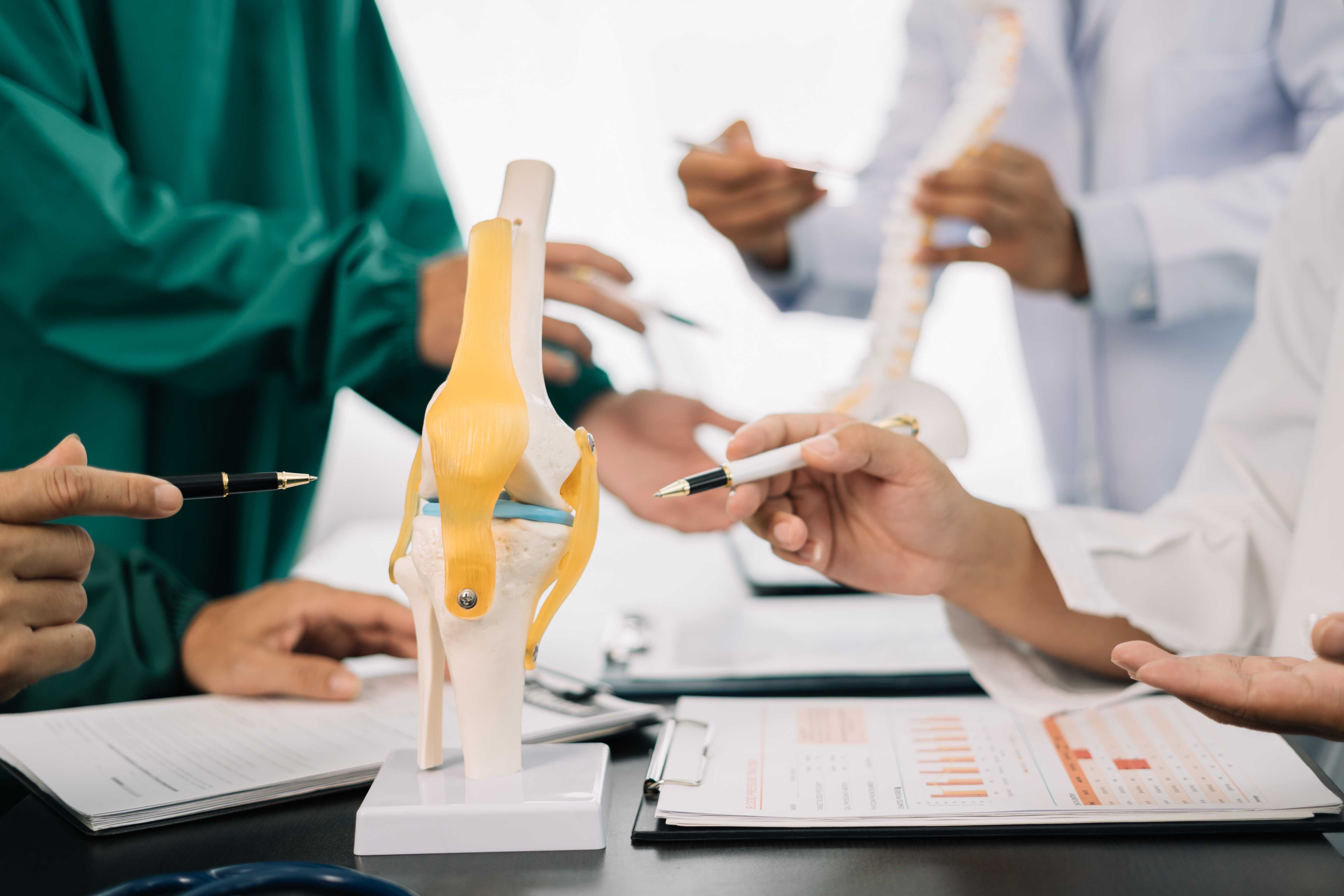
The knee joint connects the femur (thigh bone) to the tibia (shin bone). Four major ligaments hold these bones together and provide stability:
- ACL (Anterior Cruciate Ligament): Prevents the tibia from sliding forward on the femur. Controls rotational stability.
- PCL (Posterior Cruciate Ligament): Prevents the tibia from sliding backward.
- MCL (Medial Collateral Ligament): Stabilises the inner side.
- LCL (Lateral Collateral Ligament): Stabilises the outer side.
The ACL runs diagonally through the centre of the knee, from the back of the femur to the front of the tibia. During activities that involve pivoting, cutting, jumping, or sudden deceleration, the ACL bears tremendous force. When that force exceeds the ligament's tensile strength, it tears.
The critical fact that shapes the entire management of ACL tears: the ACL has virtually no capacity to heal itself. Unlike bone, which has an excellent blood supply and heals readily, the ACL is a poor healing tissue. Partial tears in specific locations may heal with time, but complete tears do not; the torn ends retract and scar tissue forms, but not functional ligament tissue. This is why surgery is recommended for most active patients with complete ACL tears.
How ACL Tears Happen
ACL tears usually occur during sudden twisting movements, awkward landings, rapid direction changes, or direct impact to the knee during sports activities.
1. Non-Contact Mechanisms (Most Common)
Approximately 70% of ACL tears are non-contact ligament tears without any direct blow to the knee.
The classic mechanism: the athlete decelerates suddenly, plants the foot, and pivots. The thigh continues rotating while the tibia is planted, creating a shear force that ruptures the ACL. This happens in:
- Cricket: A batsman diving for a run and twisting at the crease. A fielder changing direction suddenly.
- Football and futsal: A pivot to beat a defender.
- Badminton and squash: Explosive lateral movement and lunging.
- Basketball: Landing from a jump on a slightly bent, internally rotated knee.
- Gym: A misstep off a box, a squat with poor form, and a knee collapse.
2. Contact Mechanisms
Direct blows to the knee, such as a tackle in football, a collision in cricket, can also rupture the ACL, often in combination with other structures.
3. Why Women Are at Higher Risk
Female athletes tear their ACLs 2–8 times more frequently than male athletes in the same sport. The reasons are multifactorial:
- A wider pelvis creates a greater Q-angle (the angle from the hip to the knee), increasing valgus (knock-knee) forces on the ACL.
- Hormonal influences on ligament laxity.
- Neuromuscular patterns, women tend to land with less knee flexion and more valgus collapse.
- Preventable with neuromuscular training programmes (FIFA 11+, ACL prevention protocols).
Symptoms - What An ACL Tear Feels Like
At the moment of injury:
- A sudden "pop" sound or sensation described by most patients as the most distinctive feature.
- Immediate pain, though often not as severe as expected.
- A feeling that the knee has "given out" or buckled.
- Inability to continue playing.
Within the next 2–6 hours:
- Rapid swelling of the knee fills with blood (haemarthrosis). This is the most reliable early indicator of a significant intra-articular injury.
- Increasing pain and stiffness as swelling develops.
- Inability to bear full weight comfortably.
Over the following days:
- The acute pain and swelling have reduced, but the knee feels unstable.
- Difficulty with pivoting, twisting, and quick direction changes.
- Some patients can walk and even jog on an ACL-deficient knee, but the knee "gives way" with any rotational stress.
Important: Not all ACL tears produce all of these symptoms. Some patients, particularly with partial tears, have less dramatic presentations. The degree of swelling is the most reliable clinical indicator that something significant has occurred.
Grading ACL Injuries
Grade 1 (Sprain): Ligament fibres stretched but structurally intact. The knee is stable. Tenderness present, but function preserved.
Grade 2 (Partial Tear): Significant partial disruption of ligament fibres. Some laxity on testing. Variable clinical presentation, some patients have significant instability, others function reasonably well.
Grade 3 (Complete Tear): Full rupture of the ligament. Clear laxity on examination. The knee is mechanically unstable. Surgical reconstruction is typically recommended for active patients.
Diagnosis - Getting It Right
1. Clinical Examination
An experienced orthopedic surgeon can often diagnose an ACL tear from examination alone, particularly in the acute setting.
Key clinical tests:
- Lachman Test: With the knee at 30 degrees of flexion, the tibia is pulled forward. Increased forward translation compared to the other knee, positive test. The most sensitive clinical test for ACL tear.
- Anterior Drawer Test: With the knee at 90 degrees, the tibia is pulled forward. Also assesses ACL integrity.
- Pivot Shift Test: A more complex test assessing rotational instability, the hallmark of functional ACL deficiency. When positive, it produces a visible "jerk" of the tibia.
In the acute setting (first 48–72 hours), muscle spasm and pain may make examination difficult. Examination under ice or after adequate pain control is more reliable.
2. MRI Scan

This image shows a modern MRI scanner used to diagnose knee ligament injuries such as ACL and PCL tears. MRI scans provide detailed views of soft tissues for accurate injury assessment.
MRI is the definitive diagnostic investigation. It:
- Confirms the ACL tear and identifies whether it is partial or complete.
- Assesses for associated injuries, such as meniscal tears (present in 50% of ACL injuries), cartilage damage, bone bruising, MCL injury, and PCL injury.
- Guides surgical planning; the information from MRI affects graft choice, technique, and whether concurrent procedures (meniscal repair) are needed.
Every patient with a suspected ACL tear should have an MRI before any surgical decision is made.
3. X-Ray
Done initially to rule out fractures. An ACL tear may be associated with an avulsion fracture (Segond fracture, a small bony fragment off the lateral tibial plateau), which is a specific X-ray marker of ACL injury.
Treatment - Surgery Vs. Conservative Management
The surgical decision is not uniform; it depends on the patient's age, activity level, the degree of instability, and associated injuries.
1. Conservative Management (Non-Surgical)
Who is suitable for non-surgical management?
- Elderly or sedentary patients who do not participate in pivoting sports.
- Low-demand patients (Grade 3 tear but no instability symptoms in daily life).
- Patients with significant medical comorbidities that increase surgical risk.
- Patients who are willing to modify their activity level permanently (avoid pivoting sports).
What does conservative treatment involve?
- Initial RICE protocol and physiotherapy for pain and swelling management.
- Progressive quadriceps and hamstring strengthening.
- Neuromuscular training teaches the knee to compensate for ACL deficiency through muscle control.
- Activity modification, avoiding cutting and pivoting activities.
- A functional brace for support during activity.
The limitation: Even with excellent rehabilitation, a significant proportion of patients with ACL deficiency who attempt to return to cutting/pivoting sports experience ongoing instability and are at high risk for secondary meniscal and cartilage damage, which leads to accelerated arthritis. This is why surgery is recommended for most active patients.
2. Surgical Treatment: ACL Reconstruction
ACL reconstruction replaces the torn ligament with a graft, a strip of tendon from another location, that is fixed into bone tunnels to replicate the original ACL's position and function.
Graft Options
Hamstring Tendon Autograft (semitendinosus and gracilis): The most commonly used graft in India. Harvested from the back of the thigh. Excellent biomechanical properties, minimal donor site morbidity. Smaller incision than the patellar tendon. Some concern about hamstring strength reduction post-harvest, though most patients regain full strength.
Bone-Patellar Tendon-Bone (BPTB) Autograft: Uses the central third of the patellar tendon with a bone plug at each end. Considered the "gold standard" for athletes requiring the highest stability (contact sports, football). Bone-to-bone healing in the tunnels is faster. Donor site concerns: anterior knee pain ("kneeling pain"), slight risk of patellar fracture.
Quadriceps Tendon Autograft: Growing in popularity. Large cross-sectional area, excellent mechanical properties. Becoming the preferred graft in some centres for revision surgery and large patients.
Allograft (cadaver tissue): Donor tissue. Avoids donor site morbidity. Longer incorporation time. Higher re-tear rate in young, active patients is generally not the first choice for young athletes.
The Surgical Procedure: Done arthroscopically through 2–3 small incisions (< 1 cm each) using a camera and small instruments. The torn ACL remnant is removed, bone tunnels are drilled in the femur and tibia at the anatomical ACL attachment points, the graft is pulled through and fixed with titanium screws or buttons. The entire procedure takes 60–90 minutes. Hospital stay: 1–2 days.
Concurrent Procedures: If the MRI shows a meniscal tear alongside the ACL tear (very common), the meniscal repair or partial meniscectomy is performed in the same operation, fixing both problems at once.
Recovery After ACL Reconstruction - A Realistic Timeline
The 6–9 month timeline is not an industry estimate; it is based on graft biology. The graft goes through a process called ligamentisation, being gradually replaced by the body's own ACL-like tissue. Until this process is substantially complete, the graft is at its most vulnerable. Returning to sport too early dramatically increases re-tear risk.
Week 1–2 (Hospital and Home): Pain and swelling management. Straight leg raises, quad sets. Walk with crutches. Range of motion exercises begin.
Week 2–4: Transition off crutches. Range of motion improves to near full. Cycling on a stationary bike begins.
Month 1–2: Full range of motion achieved. Progressive strengthening of quadriceps, hamstrings, and hip abductors. Proprioception training begins.
Month 2–4: Running on a treadmill begins (straight line only). Progressive loading leg press, step-ups, lunges. Swelling should be minimal at rest.
Month 4–6: Jogging transitions to running. Sport-specific movement begins with cutting, jumping, and pivoting, but not in competitive situations. Return-to-sport testing begins.
Month 6–9: Gradual return to full training and then competitive play guided by functional testing (limb symmetry index, hop tests) rather than time alone.
Re-tear risk: The overall re-tear rate for ACL reconstruction is approximately 5–10% in the general patient population. In young athletes under 25 who return to high-demand sport, it is higher. This is why the return-to-sport timeline, proper rehabilitation, and neuromuscular training for landing mechanics are all critical.
ACL Injury And Long-Term Knee Health
An important, often underemphasised point: ACL injury, even when surgically reconstructed, is associated with an increased long-term risk of knee osteoarthritis. This risk is partly from the injury itself (bone bruising, associated cartilage and meniscal damage) and partly from the altered biomechanics of the reconstructed joint.
Reducing long-term arthritis risk after ACL reconstruction involves:
- Maintaining a healthy body weight (reduces joint load).
- Continuing with low-impact exercise long-term (swimming, cycling).
- Addressing any associated meniscal pathology at the time of surgery rather than leaving it untreated.
- Committing to lifelong quadriceps and hamstring strength maintenance.
ACL Treatment In Noida - Dr. Mayank Chauhan At Prakash Hospital
Dr. Mayank Chauhan, Senior Orthopedic Surgeon at Prakash Hospital, Sector 33, Noida, manages ACL injuries from initial evaluation through arthroscopic reconstruction and return-to-sport rehabilitation. For patients in Noida, Greater Noida, and Delhi NCR who have sustained a knee injury with swelling and instability, early consultation with appropriate MRI imaging will establish exactly what has been injured and what the best management path is.
Not every ACL tear requires immediate surgery. And not every patient who thinks they have an ACL tear actually does. Getting the right diagnosis first is everything. To book a consultation, call the number listed on the website.
The Bottom Line

A doctor is helping a female patient wearing a knee brace in physiotherapy.
An ACL tear is a serious injury, but it is an injury that, with proper diagnosis, appropriate surgical management for active patients, and committed rehabilitation, most people recover fully. The majority of patients who undergo ACL reconstruction and complete their rehabilitation return to sport at the same level or above. The keys: accurate diagnosis, choosing the right graft with your surgeon, trusting the timeline, and not rushing return to sport.
To consult Dr. Mayank Chauhan, Senior Orthopedic Surgeon in Noida, call the number listed on the website.