Scoliosis in Adults — Causes, Symptoms, and When Treatment Is Needed

The image shows a woman holding her lower back with a highlighted spine, indicating pain or spinal discomfort. It visually represents back pain commonly associated with spinal conditions such as scoliosis or poor posture.
Most people associate scoliosis with school-age children and the awkward posture check in PE class. The diagnosis, in that context, is about monitoring a sideways curve in the spine during growth — and in many cases, it causes no long-term problems.
Adult scoliosis is a different condition. It is managed differently, causes different symptoms, and the decisions around treatment involve a very different set of considerations.
In adults, scoliosis either represents a childhood curve that was never treated and is now causing problems in middle age, or — increasingly commonly — a newly developed degenerative curve driven by disc degeneration, osteoporosis, and asymmetric collapse of the aging spine.
Both types affect quality of life. Both deserve proper assessment.
What Is Scoliosis?
Scoliosis is defined as a lateral (sideways) curvature of the spine with a Cobb angle of 10 degrees or more. The Cobb angle is measured on a standing X-ray by drawing lines along the top and bottom of the most tilted vertebrae at each end of the curve.
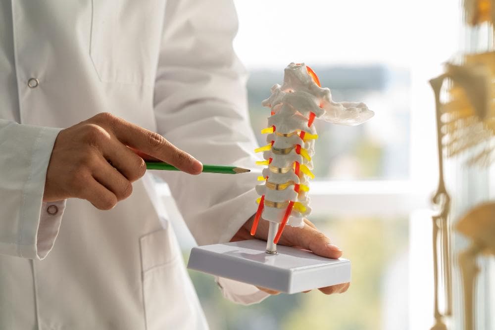
The spine is normally straight when viewed from the front, and has natural curves front-to-back (lordosis in the lumbar spine, kyphosis in the thoracic spine). In scoliosis, the spine bends side-to-side in an S or C shape, often with associated rotation of the vertebrae — a three-dimensional deformity, not just a simple tilt.
Types of Adult Scoliosis
Idiopathic Scoliosis in Adults (AIS Continuing into Adulthood)
Adolescent idiopathic scoliosis (AIS) — the most common form in young people — develops during growth spurts and has no known cause (hence "idiopathic"). In most adolescents, curves are mild and cause no long-term problems.
In a proportion, curves are larger (over 40–50 degrees) and progress slowly even after skeletal maturity. These patients reach adulthood with a structural curve that may worsen by 0.5–1 degree per year and cause progressive pain, cosmetic concern, and occasionally, respiratory compromise if thoracic curves are large.
Degenerative Scoliosis (De Novo Scoliosis)
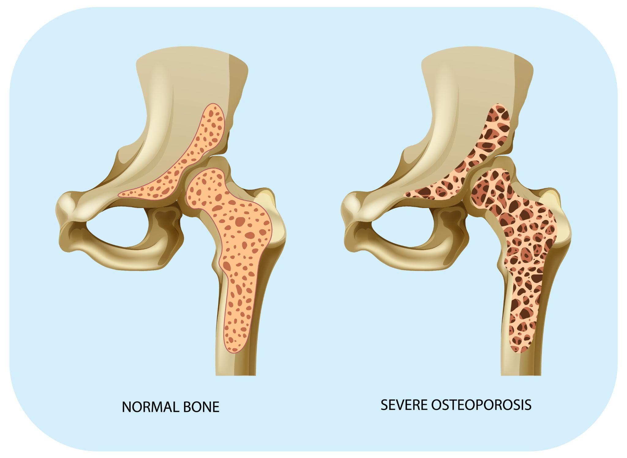
The form increasingly seen in adults over 50. As discs degenerate asymmetrically — losing height and collapsing more on one side than the other — and as osteoporotic vertebral compression fractures compress one side of a vertebra, the spine gradually bends laterally.
This process is distinct from idiopathic scoliosis and develops entirely in adulthood. It is the most common form of scoliosis seen in Indian adults at orthopedic and spine clinics.
Key features of degenerative scoliosis:
- Lumbar spine is most commonly affected
- The curve develops gradually — many patients don't notice it until it becomes significant
- Pain is often the primary complaint, unlike in adolescent scoliosis which is typically painless
- The collapsing discs and facet joints also narrow the spinal canal and nerve exit foramina, causing leg pain, numbness, and weakness (neurogenic claudication) — symptoms indistinguishable from spinal stenosis, which often coexists
Symptoms of Adult Scoliosis
Adult scoliosis causes a different symptom profile from its adolescent counterpart:
Back pain: The most consistent complaint. Unlike adolescent scoliosis, degenerative adult scoliosis is often painful. The asymmetric loading of facet joints, the paraspinal muscle fatigue from working against the imbalanced curve, and the disc degeneration itself all contribute. Pain is typically worse with prolonged standing or walking, and relieved by sitting or lying down.
Radicular leg pain: When foraminal stenosis (narrowing of nerve exit channels) develops alongside the curve, patients experience shooting pain, tingling, or weakness radiating down one or both legs. Many patients notice they can walk only a short distance before leg pain forces them to stop and sit down (neurogenic claudication).
Visible asymmetry: Uneven shoulders, uneven hip heights, visible waistline asymmetry, a prominent rib hump (usually in thoracic curves). Many patients notice their posture has changed and they cannot stand fully straight.
Height loss: Significant degenerative scoliosis combined with vertebral compression fractures can cause measurable height loss over years.
Difficulty standing erect: Some patients with severe lumbar degenerative scoliosis cannot straighten up fully. This is called sagittal imbalance — the spine tips forward, and patients compensate by flexing their hips and knees.
Diagnosis
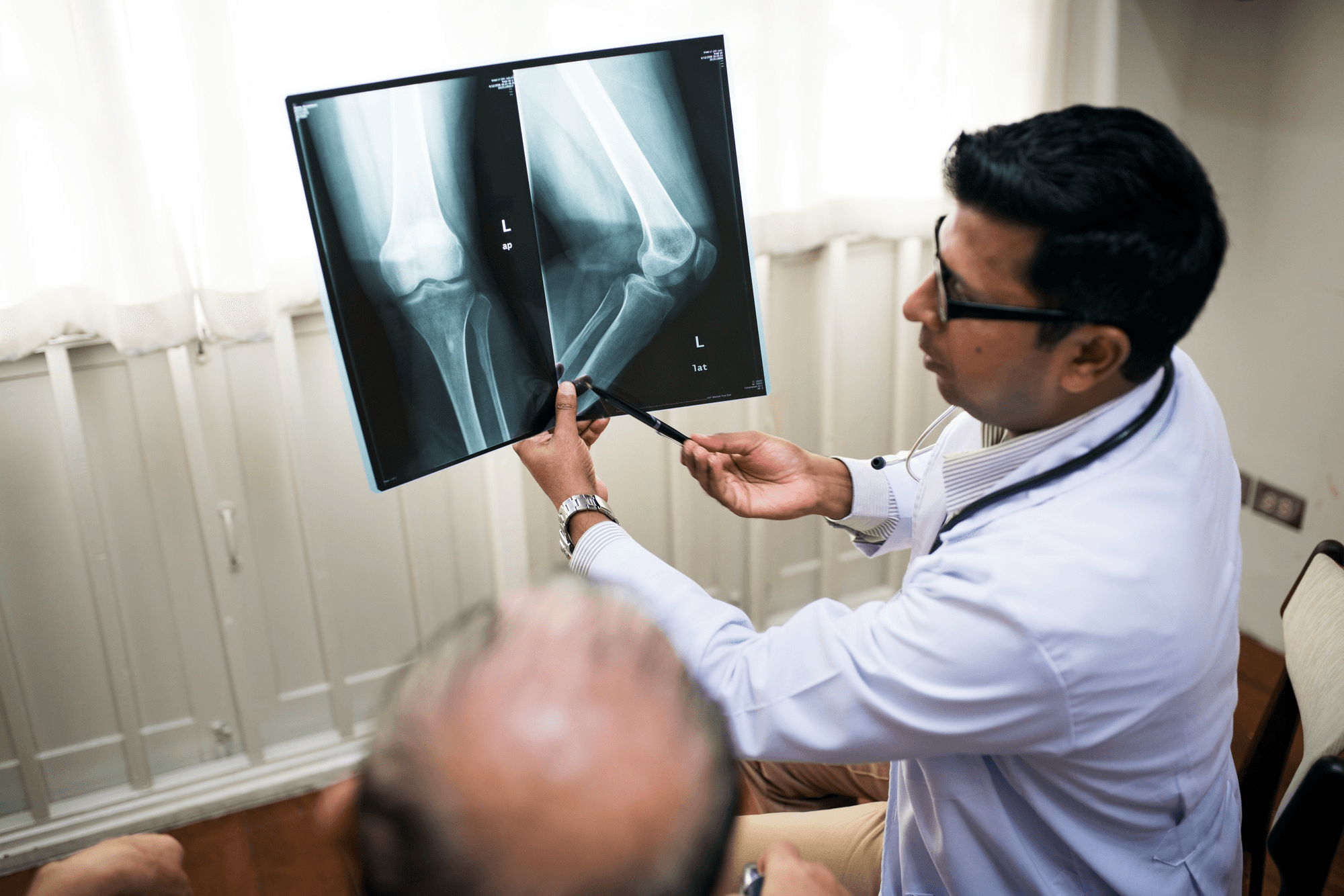
Standing full-spine X-rays: The essential investigation. X-rays are taken standing, in the same position every time for serial comparison. The Cobb angle is measured. Alignment in both the coronal (front-to-back) and sagittal (side-to-side) planes is assessed.
MRI: When nerve compression symptoms are present — leg pain, numbness, weakness — MRI is required to assess disc herniations, foraminal stenosis, and spinal cord or nerve root compression.
CT scan: For detailed bony anatomy before surgical planning, and for assessment of bone quality.
DEXA scan: In older patients, bone density assessment is important because osteoporosis significantly affects both the natural history of the curve and surgical planning (poor bone quality complicates instrumentation).
Treatment: Non-Surgical First
The large majority of adult scoliosis patients do not need surgery. The goal is symptom management and preventing unnecessary functional decline.
Physiotherapy
Core strengthening — particularly the paraspinal muscles that support the spine — reduces the muscle fatigue that causes pain in scoliosis. It cannot straighten the curve, but it can meaningfully reduce pain and improve function.
Specific approaches:
- Schroth therapy: A breathing and exercise method specifically designed for scoliosis. Involves specific postures and breathing patterns to work against the curve's direction. Growing evidence base for pain reduction and posture improvement in adults.
- Core stabilisation exercises: Targeting the multifidus, transversus abdominis, and gluteal muscles that provide spinal stability.
- Aquatic physiotherapy: Reduces load on the spine while allowing muscle activation.
Pain Management
- NSAIDs for pain and inflammation
- Muscle relaxants for paraspinal muscle spasm
- Nerve pain medications (gabapentin, pregabalin) when radicular leg symptoms are present
- Epidural steroid injections or facet joint injections: Guided injections for pain relief, particularly for leg symptoms. Can provide meaningful short-to-medium term relief, serving as a bridge to physiotherapy or deferring surgery.
Bracing
In adult scoliosis, bracing plays a different and more limited role than in adolescent scoliosis. Braces do not straighten an established adult curve. They can provide postural support and pain relief in selected patients (particularly those with lumbar degenerative scoliosis who notice postural improvement with a lumbar support). They are generally not used as a permanent solution.
Surgery: When Is It Needed?
Only a minority of adult scoliosis patients need surgery. The indications are specific:
- Curve greater than 50–55 degrees that is documented to be progressively worsening
- Severe pain unresponsive to 6+ months of appropriate conservative management
- Progressive neurological deficit (worsening weakness, sensory loss, bladder/bowel involvement)
- Significant sagittal imbalance causing inability to stand erect and limiting daily function
- Cosmetic concerns sufficient to significantly impact quality of life (less commonly the primary indication in adults)
The surgery for adult degenerative scoliosis is major. It typically involves long-segment spinal fusion — connecting many vertebrae together with rods, screws, and bone graft to straighten and stabilise the curve. In many cases, decompression (removal of bone or disc material compressing nerves) is performed simultaneously.
Recovery is prolonged (6–12 months to full recovery), and complications are more common than in simpler spinal procedures. The decision to operate requires careful discussion of realistic expectations, surgical risk, and alternatives.
Minimally invasive approaches to scoliosis correction (percutaneous screw placement, lateral interbody fusion) have evolved significantly and are available at specialist centres in India. They reduce muscle trauma compared to traditional open approaches.
Spine Assessment in Noida
Dr. Mayank Chauhan at Prakash Hospital, Sector 33, Noida, evaluates adult spinal deformity, including scoliosis, as part of comprehensive spine care. He assesses symptom severity, curve magnitude, and the presence of nerve compression before recommending the appropriate level of intervention — conservative management, injection procedures, or referral for surgical evaluation in complex cases.
To book a consultation, call the number listed on the website.