How Bone Fractures Heal — The 4 Stages Explained
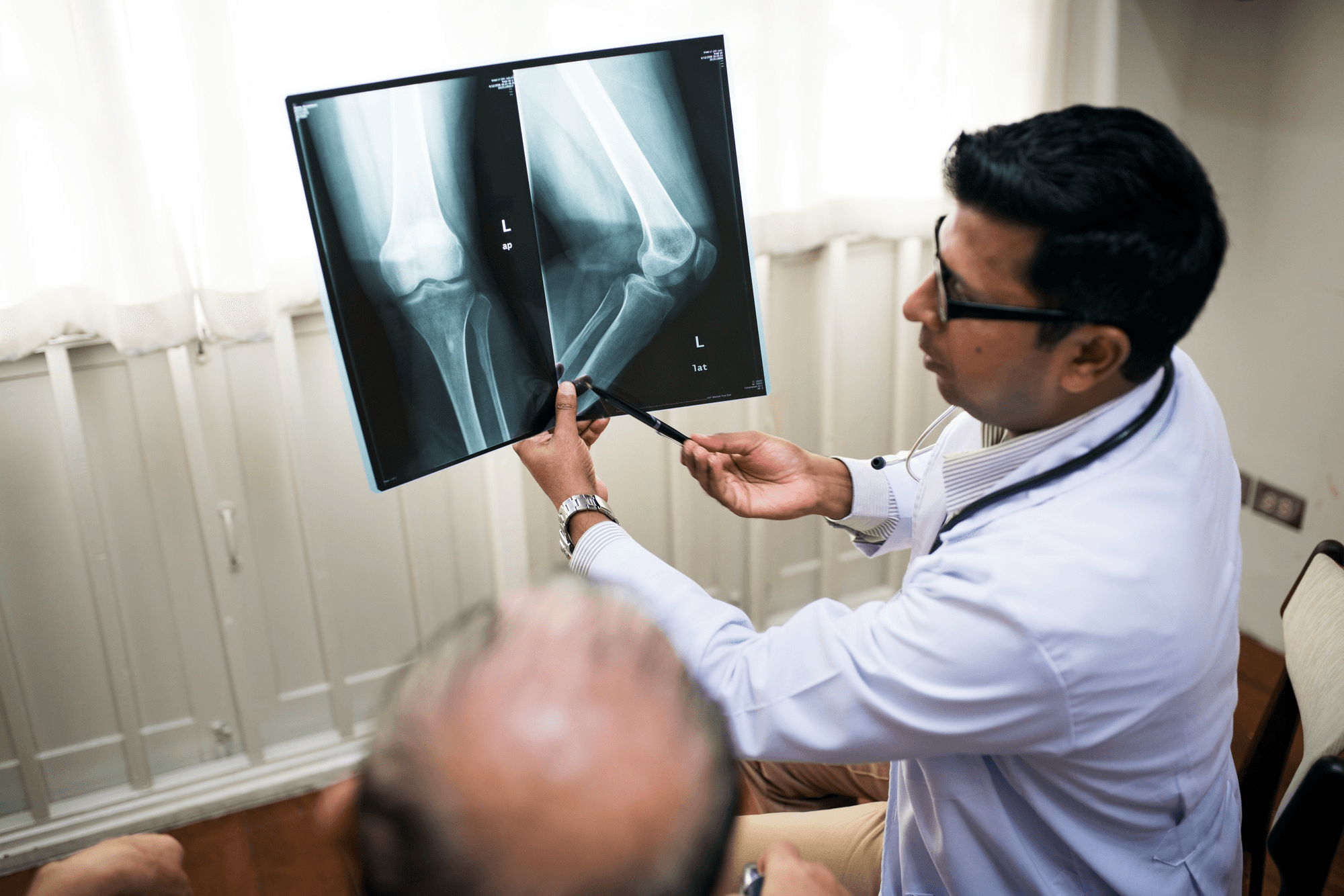
X-ray imaging test used to diagnose bone fractures and detect structural abnormalities.
Most patients who break a bone receive a plaster cast or a surgical implant, are told to follow up in 4–6 weeks, and leave the appointment with a general sense that the bone will "knit back together" — but with no real understanding of how.
That understanding matters. Knowing what's happening inside the bone during healing helps patients understand why activity restriction matters, why nutrition matters, why smoking slows healing, and why their doctor is making specific decisions at specific time points.
Bone fracture healing is one of the more extraordinary biological processes in the body — a complete regeneration of mineralised tissue from injury to strength restoration.
The Biology of Bone: Why Fractures Can Heal
Unlike most soft tissues, bone has a remarkable capacity to heal with true regeneration — meaning the healed bone is structurally equivalent to the original, not a weaker scar tissue substitute.
This is possible because bone is living tissue, containing:
- Osteoblasts: Cells that build new bone by secreting collagen matrix and directing mineralisation
- Osteoclasts: Cells that resorb old or damaged bone
- Osteocytes: Embedded bone cells that sense mechanical load and regulate remodelling
- Periosteum: The fibrous layer covering bone's surface, rich in stem cells that participate in fracture repair
When a bone breaks, all of these players mobilise as part of an organised, precisely timed repair sequence.
Stage 1: Haematoma Formation (Days 1–5)
The first event after a fracture is bleeding. Blood vessels within the bone (Haversian canals) and in the surrounding soft tissue are torn by the fracture. Blood accumulates at the fracture site, forming a haematoma — a clot.
This is not just bleeding. The haematoma is the scaffold on which all subsequent healing is built. It contains growth factors that signal the beginning of repair:
- Platelet-derived growth factor (PDGF): Stimulates stem cell migration to the fracture site
- Transforming growth factor-beta (TGF-β): Drives both cartilage and bone formation
- Vascular endothelial growth factor (VEGF): Promotes new blood vessel formation (angiogenesis) into the avascular clot
Inflammation is prominent in Stage 1. The area is swollen, warm, red, and painful. This is not a complication — it is the required biological response. Anti-inflammatory medications given in very high doses in the first days of fracture can actually impair healing by suppressing this inflammatory signal. This is why NSAIDs are used cautiously in the early fracture period.
Stage 2: Soft Callus Formation (Days 5–21)
As the haematoma organises and blood supply returns to the fracture zone, specialised cells called chondrocytes and osteoblasts begin building the bridge between fracture fragments.
The initial bridge is a soft callus — a collagen and cartilage scaffold that connects the broken ends. It has no mineral content yet and therefore no structural strength. On X-ray at this stage, the fracture gap is still visible and the callus appears as a faint, hazy shadow around the fracture.
Clinically, the bone is stable enough that the sharp pain of early fracture has subsided somewhat, but the bone has no load-bearing strength. Disrupting this phase — through excessive movement of an inadequately immobilised fracture — causes damage to the developing callus and prolongs healing.
This is why immobilisation in the first 3–6 weeks is so critical.
Stage 3: Hard (Bony) Callus Formation (Weeks 3–12)
This is the transformation phase. The soft cartilage callus progressively mineralises — calcium phosphate crystals are deposited into the collagen matrix, converting the flexible cartilage bridge into hard woven bone.
This process, called endochondral ossification (the same process by which bones develop before birth), produces a hard callus visible on X-ray as dense new bone around the fracture site.
By the end of Stage 3, the fracture is mechanically stable. In clinical terms, this is when:
- Long bone fractures can bear weight progressively (under guidance)
- Wrist fractures allow removal of the cast
- Shoulder fractures allow beginning range-of-motion exercises
The timeline varies significantly by bone:
- Wrist fracture: Hard callus forms in 4–8 weeks
- Tibial shaft fracture: 8–16 weeks
- Femoral shaft fracture: 12–24 weeks
- Ribs: 4–6 weeks
- Vertebral compression fracture: 6–12 weeks
These timelines are influenced by the patient's age, bone quality, fracture location and type, fixation method, and nutritional status.
Stage 4: Remodelling (Months to Years)
The hard callus formed in Stage 3 is woven bone — structurally strong but architecturally disorganised. It forms a bulge visible on X-ray around the fracture site.
Stage 4 is a prolonged refinement process. Osteoclasts resorb excess or disorganised bone. Osteoblasts deposit new, architecturally aligned lamellar bone along the lines of mechanical stress. The fracture callus is gradually replaced by bone that is structurally equivalent to the original.
Over 1–2 years, the X-ray appearance of the healed fracture fades as the remodelled bone more closely resembles normal bone. In children, this process is so complete that the fracture site may be invisible on X-ray. In adults, a faint shadow often remains.
This stage is why bone looks fully healed on X-ray before the bone has regained full structural strength, and why physiotherapy and progressive loading are important throughout.
What Affects the Speed of Healing
Age
Children heal fractures significantly faster than adults. The periosteum is thicker and more active, the blood supply is richer, and the growth plates contribute to rapid repair. A 6-year-old with a femoral fracture may walk within 3–4 weeks. A 70-year-old with the same fracture will take significantly longer.
As adults age beyond 50, healing slows further due to reduced osteoblast activity and, often, concurrent osteoporosis.
Bone Quality (Osteoporosis)
Osteoporotic bone has reduced mineral density and thinner cortices. Fractures in osteoporotic bone heal, but may take longer, carry greater implant failure risk, and can collapse further during healing. Treating underlying osteoporosis both before elective orthopedic surgery and after fragility fractures is important.
Fracture Pattern
Simple transverse fractures with good bony contact heal faster than comminuted (multi-fragment) fractures with large gaps. Fractures with extensive soft tissue damage around them have impaired blood supply to the repair zone.
Blood Supply
Bones with good blood supply heal faster. The femoral neck has particularly precarious blood supply — which is why femoral neck fractures in young adults are treated urgently (to preserve the blood supply before avascular necrosis develops) and why healing is unreliable enough that hip replacement is often preferred over fixation in older patients.
Infection
Infection dramatically impairs fracture healing. Bacteria release enzymes that break down the repair matrix and compete for nutrients at the healing site. Open fractures (where bacteria have entered the wound) require meticulous wound management and antibiotics.
Nutrition
Adequate protein (1.2–1.5 g/kg/day), calcium (1,000–1,200 mg daily), vitamin D (essential for calcium absorption), vitamin C (for collagen synthesis), and zinc (for cell division and tissue repair) all support fracture healing. Nutritional deficiency prolongs healing.
Smoking
Nicotine constricts blood vessels supplying the healing fracture, reducing oxygen delivery to a tissue that requires high oxygen levels for osteoblast activity. Studies show fractures in smokers take 25–50% longer to heal, and non-union rates are significantly higher.
Fixation Quality
Well-aligned fractures heal faster than malaligned ones. Fractures with secure fixation (providing enough stability for the callus to form without disruption) heal faster than unstable ones.
When Fractures Don't Heal: Non-Union and Delayed Union
Delayed union: Healing is taking longer than expected but is still progressing. Usually managed with observation, nutritional optimisation, and if needed, additional intervention.
Non-union: The fracture has stopped healing — there is no evidence of progressive callus formation on serial X-rays over 6 months. The fracture ends may become rounded and sclerotic (hard and dense) as the body treats the gap as a pseudo-joint rather than a healing fracture.
Causes of non-union include inadequate immobilisation, infection, poor blood supply, large fracture gaps, systemic factors (diabetes, vitamin D deficiency, smoking), and certain medications (bisphosphonates can rarely cause atypical fractures and impair healing).
Treatment of non-union may involve:
- Surgical revision of fixation
- Bone grafting (adding bone material to stimulate repair)
- Electrical stimulation of bone healing
- PRP (platelet-rich plasma) injection into the fracture site
- Correction of underlying nutritional or systemic factors
Fracture Care at Prakash Hospital, Noida
Prakash Hospital at Sector 33, Noida, has 24/7 trauma and orthopedic care. Dr. Mayank Chauhan manages both acute fracture fixation and complex non-unions requiring revision surgery. The hospital is empanelled with CGHS, ECHS, ESI, and all major private insurers.
To book a consultation or visit the emergency department, call the number listed on the website.