Tendon Injuries — Types, Causes, and Treatment From Conservative to Surgery
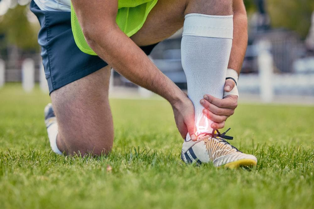
A male soccer player kneels on the grass, gripping his ankle with both hands and showing a pained expression. The area around his ankle is highlighted in red, indicating injury. The background features a blurred field and goalposts.
Tendons are among the strongest structures in the body — dense, fibrous cords that connect muscle to bone, transmitting the enormous forces generated by muscle contraction into movement. The Achilles tendon, for example, withstands forces of up to 12 times body weight during running.
And yet tendons are also among the most commonly injured structures in orthopedic medicine. This paradox has a straightforward explanation: tendons are poorly vascularised (relatively little blood supply), which means they heal slowly, respond poorly to chronic overload, and when they fail catastrophically, the failure is often dramatic.
Understanding the different types of tendon injury — and the distinct management approaches for each — is genuinely useful for anyone active in sport, anyone with chronic joint pain, or anyone who has been told they have a "tendon problem" without further explanation.
The Three Types of Tendon Injury
1. Tendinopathy (Chronic Tendon Degeneration)
The most common presentation. Not an acute injury, not truly inflammatory in the classic sense — tendinopathy is a chronic degenerative process within the tendon structure.
The pathology involves disordered collagen fibre arrangement, increased ground substance, abnormal blood vessel ingrowth (neovascularisation), and reduced tensile strength — all without the classical inflammatory infiltrate that the older term "tendinitis" implied. This is why tendinopathy is a more accurate term than tendinitis for most chronic cases.
Causes: Repetitive loading beyond the tendon's capacity for adaptation. Usually a combination of volume, intensity, and inadequate recovery. Common precipitating factors: sudden increase in training load, change in footwear or surface, return to sport after a break, poor biomechanics.
Presentation: Gradual onset of pain over and around the tendon insertion or mid-portion. Characteristically worse with initial loading (first steps in the morning), improving with light warm-up, worsening again with sustained activity. No acute event.
2. Acute Partial Tear
A sudden injury causes some fibres to rupture while others remain intact. This is distinct from tendinopathy in that there is a specific moment of injury — typically a sudden eccentric load (the muscle contracting while the joint is extending).
Presentation: Sudden pain at the time of injury. Localised swelling and bruising within 24–48 hours. Tenderness directly over the tear. Some preserved function — the tendon hasn't completely failed, but activity is painful and force-generating capacity is reduced.
3. Complete Rupture
All or nearly all fibres are torn. Catastrophic mechanical failure of the tendon. Often preceded by chronic degeneration, making the tendon structurally weaker before the final rupture occurs.
Presentation: Sudden, severe pain. Often an audible "pop." Immediate loss of function — for Achilles rupture, inability to plantarflex the foot; for quadriceps tendon rupture, inability to extend the knee. A palpable gap in the tendon may be felt.
The Major Tendons That Fail
Achilles Tendon
The most commonly ruptured tendon in the body. The Achilles connects the calf muscles (gastrocnemius and soleus) to the heel bone. Ruptures typically occur 2–6 cm above the heel insertion — a zone of relatively poor blood supply and common degenerative change.
Who is at risk: Men aged 30–50 who are active in recreational sport (badminton, basketball, squash, cricket — any sport with explosive push-off). Often called a "weekend warrior" injury.
The Thompson test: Squeezing the calf with the patient lying face down. In a healthy Achilles, the foot plantarflexes. In a complete rupture, the foot remains still.
Treatment — the historic debate: For decades, Achilles rupture was treated surgically (open repair of the torn ends) or non-surgically (casting in equinus position for 6–8 weeks). Large randomised trials have shown that functional non-surgical treatment using early weight-bearing in a walking boot produces equivalent re-rupture rates and functional outcomes to surgery in the general population. This has shifted practice — non-surgical management with early weight-bearing is now the standard for many patients.
Surgery is preferred for elite athletes, certain high-risk tear patterns (large gaps, complex tears), and active patients who want the security of surgical repair.
Chronic Achilles tendinopathy: Pain in the Achilles tendon without acute rupture, treated with eccentric calf raises (the Alfredson protocol remains the best-evidenced rehabilitation exercise for mid-portion Achilles tendinopathy), activity modification, and if conservative management fails, shockwave therapy or PRP injection.
Patellar Tendon (Jumper's Knee)
The patellar tendon connects the kneecap (patella) to the tibial tuberosity (the bump at the top of the shin). It transmits the quadriceps force to extend the knee.
Patellar tendinopathy — "jumper's knee" — is extremely common in volleyball, basketball, kabaddi, and any sport involving repeated jumping. Pain is at the inferior pole of the patella (just below the kneecap) and is aggravated by jumping, landing, squatting, and stairs.
Treatment: Eccentric quadriceps exercises (slow decline squats on a 25-degree board) are the cornerstone of evidence-based management. Progress through loading volumes rather than treating with rest alone. PRP injection is increasingly used for refractory cases with growing evidence of benefit. Surgery is rarely needed.
Patellar tendon rupture: Complete rupture usually follows a sudden forceful deceleration or landing, often in already-degenerated tissue. Causes acute inability to extend the knee. Requires surgical repair.
Quadriceps Tendon
Above the kneecap. Rupture typically occurs in older patients (over 60) and is associated with conditions that weaken tendons: kidney disease, hyperparathyroidism, corticosteroid use. Complete ruptures require surgical repair — the quadriceps tendon is too central to knee extension to manage non-surgically.
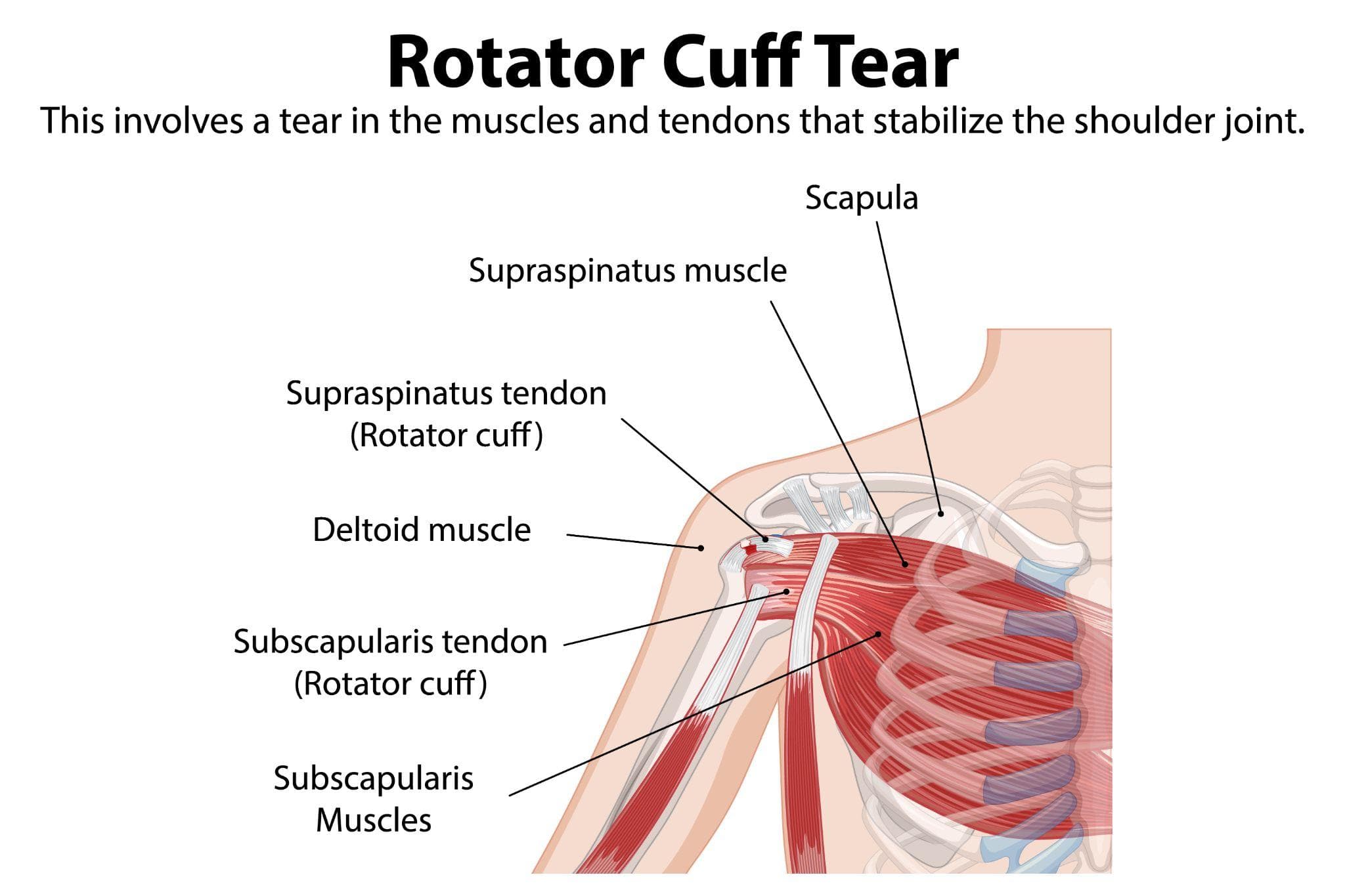
Rotator Cuff Tendons (Covered in Detail in Blog 41)
Supraspinatus is the most commonly torn rotator cuff tendon, from impingement, chronic overhead loading, or acute injury. See the rotator cuff blog for full detail.
Tibialis Posterior Tendon
Runs along the inside of the ankle, stabilising the arch and enabling foot inversion. Tibialis posterior tendon dysfunction (TPTD) is a common cause of adult-acquired flat foot deformity, particularly in women over 50 with excess weight. The tendon gradually tears and weakens, allowing the arch to collapse.
Presentation: Pain along the inner ankle and foot. Progressive flatfoot deformity. Inability to do a single-leg heel raise on the affected side. "Too many toes" sign when viewed from behind (the toes are visible lateral to the heel because of flatfoot).
Treatment: Orthotics (arch supports), physiotherapy, and sometimes immobilisation for early stages. Surgical reconstruction for advanced failure.
Common Extensor Tendons of the Elbow (Tennis Elbow)
Lateral epicondylitis ("tennis elbow") is tendinopathy of the common extensor tendon origin at the lateral epicondyle of the humerus. Despite the name, it affects computer users, manual workers, and homemakers as often as tennis players.
Treatment: Physiotherapy emphasising eccentric wrist extension exercises, activity modification, elbow straps, and if needed, corticosteroid injection (effective short-term) or PRP injection. Surgery is rarely required but can address resistant cases.
General Principles of Tendon Treatment
Conservative First
For tendinopathy (the chronic form), the research consistently supports loading — specific, graduated mechanical loading of the tendon — as the most effective treatment. Complete rest is counterproductive: tendons need load to reorganise and strengthen.
The three-phase loading approach:
- Isometric loading: Holding a contraction (e.g., single-leg wall sit for patellar tendinopathy) to reduce pain initially without movement
- Isotonic loading: Slow repetitive movements through range (e.g., slow eccentric heel drops for Achilles)
- Energy storage/sport-specific loading: Gradual return to sport-specific movement, jumping, and plyometrics
Injection Treatments
Corticosteroid: Effective for short-term pain relief in tendinopathy. Multiple injections risk tendon structural weakening and should be used sparingly — generally not more than 2–3 over a tendon's lifetime.
PRP (Platelet-Rich Plasma): Centrifuged platelet concentrate containing growth factors injected into the tendon. Growing evidence base, particularly for chronic patellar tendinopathy and chronic Achilles tendinopathy. Best evidence when used alongside structured loading rehabilitation.
Surgical Tendon Repair
For complete ruptures: surgical repair is often required to restore the structural continuity and force-transmission capacity of the tendon. The technique varies by tendon — direct end-to-end suture for Achilles, anchor-based repair for rotator cuff, suture anchor or drill-hole repair for patellar tendon.
For tendinopathy refractory to all conservative measures: surgical debridement removes degenerative tissue and stimulates a healing response. Used selectively.
Sports and Tendon Care at Prakash Hospital, Noida
Dr. Mayank Chauhan evaluates all major tendon injuries at Prakash Hospital, Sector 33, Noida — from chronic tendinopathy managed conservatively to acute ruptures requiring surgical repair. MRI is used to characterise tear extent before treatment decisions are made.
To book a consultation, call the number listed on the website.