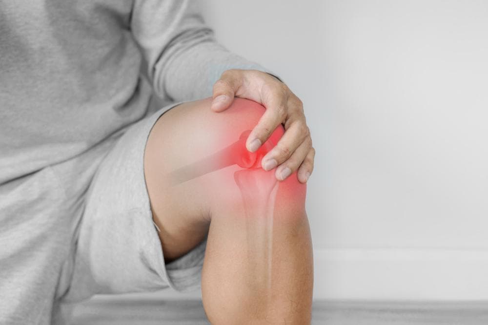
Meniscus Surgery — Repair vs Removal: What's the Difference and Which Is Better?
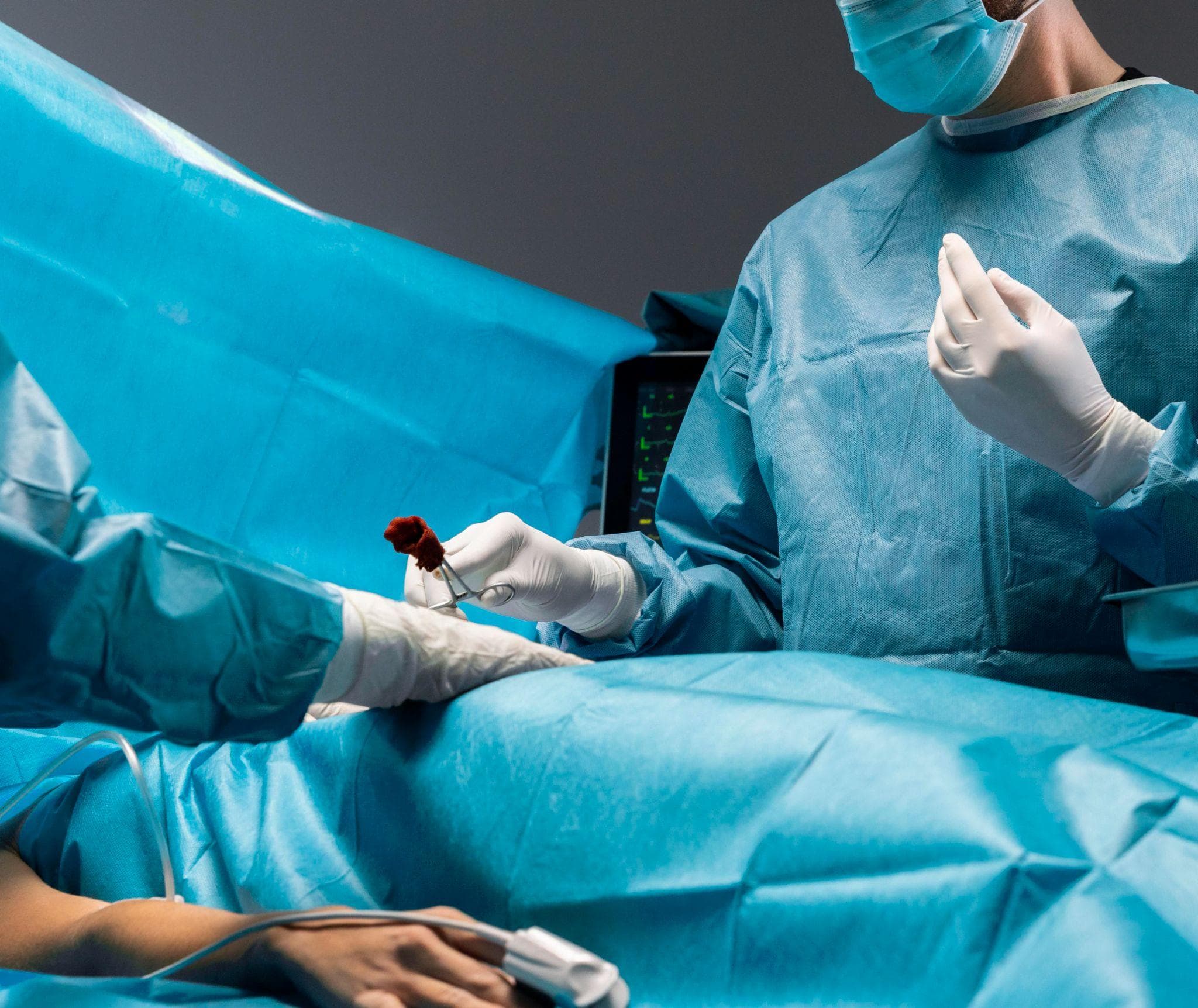
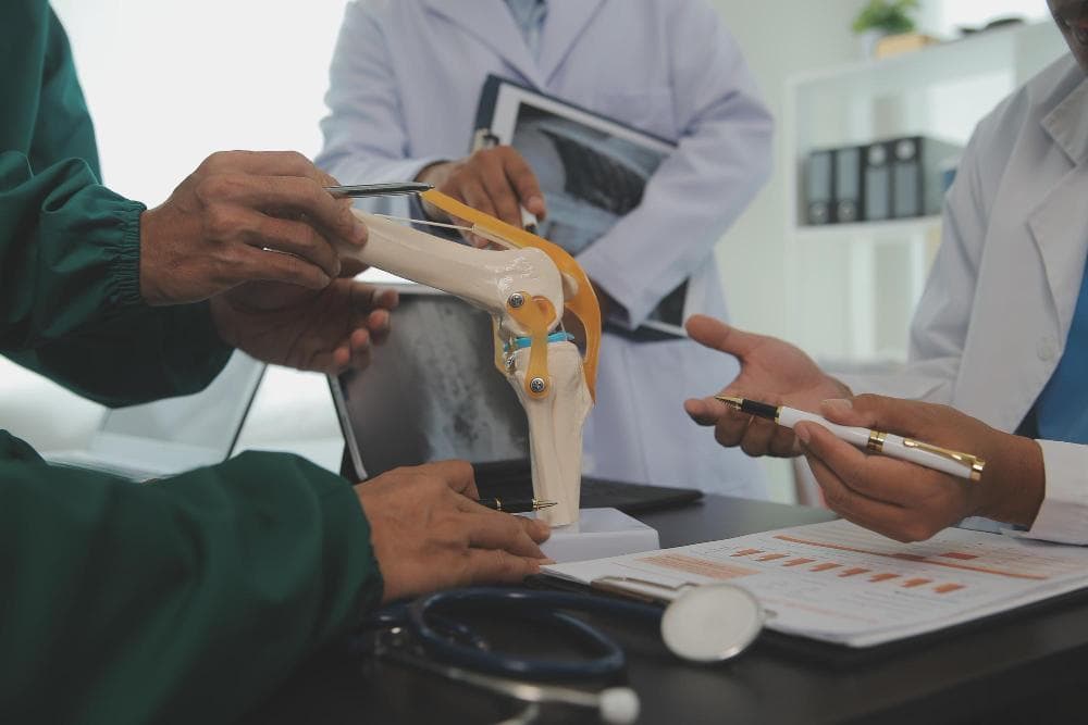
Orthopedic surgeons performing surgery.
The meniscus gets injured more often than almost any other structure in the knee. In India — where cricket, kabaddi, football, and gym training all place rotational and compressive loads on the knee — meniscal tears are among the most common orthopedic presentations seen in sports medicine clinics.
When surgery is recommended, patients almost universally ask the same question: "Will you fix it or remove it?"
The answer depends on factors that are not about preference — they're about biology, tear pattern, blood supply, and patient age. Getting this decision right has long-term implications for the health of the knee. Getting it wrong — particularly removing tissue that could have been repaired — accelerates cartilage wear and increases the lifetime risk of knee arthritis.
What the Meniscus Does
The knee has two menisci — the medial (inner) and lateral (outer). Each is a C-shaped wedge of fibrocartilage sitting between the femur and tibia. They serve several critical functions:
Load distribution: The menisci increase the contact area between the curved femoral condyle and the relatively flat tibial plateau, distributing compressive load across a larger surface area. Remove a meniscus entirely, and the load concentrates on a much smaller area of cartilage.
Shock absorption: The menisci absorb 50–70% of compressive forces during weight-bearing. After total meniscectomy, these forces are transmitted directly to the articular cartilage.
Joint stabilisation: Particularly the posterior horn of the medial meniscus, which provides secondary stability to anterior-posterior translation of the tibia.
Lubrication: The menisci help distribute synovial fluid across the joint surface.
Long-term studies consistently show that knees with significant portions of meniscus removed develop arthritis faster than those where the meniscus is preserved. This is the fundamental biological argument for repairing rather than removing wherever the biology allows it.
How Meniscal Tears Are Classified
Tears are described by their location and pattern — both of which determine whether repair is feasible.
By Location: The Zone System
The meniscus is divided into zones based on blood supply:
Red-red zone (outer third): Richly vascularised. Tears here have the best healing potential after repair. If you're going to repair any tear, it should be here.
Red-white zone (middle third): Moderately vascularised at the red side, avascular at the white side. Healing potential is intermediate. Repair is attempted for appropriate tear patterns in this zone, though healing is less reliable than the outer zone.
White-white zone (inner third): Completely avascular. No blood supply means no healing capacity. Tears in this zone cannot reliably heal after repair and are treated by removal.
By Pattern
Longitudinal tears (including bucket-handle tears): Run parallel to the meniscus circumference. Bucket-handle tears are a complete longitudinal tear through the full thickness, causing the inner portion to displace into the joint and lock the knee. Repair is often possible for acute longitudinal tears in the vascular zone.
Radial tears: Perpendicular to the circumference, disrupting the hoop stress function of the meniscus. More difficult to repair; some are treated with partial removal.
Horizontal tears: Run horizontally through the meniscus thickness, creating two leaves. Common degenerative pattern. Generally not repairable in most cases.
Complex tears: Multiple planes of tearing. Often treated with partial removal.
Degenerative tears: From wear rather than acute trauma. Typically involve the white zone and degenerate meniscal tissue. Almost always treated with removal of the unstable fragment.
Option 1: Meniscus Repair
Repair involves placing sutures through the torn meniscal tissue to hold the edges together while biology heals the tear from the inside out.
Who Is a Good Candidate for Repair?
- Tear in the red-red or red-white zone (outer or middle meniscus, where blood supply exists)
- Longitudinal or vertical tear pattern — these respond best to repair
- Younger patient (under 40–45) — better healing capacity and justification for more intensive rehabilitation
- Acute tear (recent injury, usually within 3–6 months) — chronic tears with degenerated edges don't heal as reliably
- Tear length typically under 4 cm — larger tears are more challenging to repair reliably
- Accompanying ACL reconstruction — when ACL repair is done simultaneously, the synovial reaction enhances meniscal healing. Meniscal repair with concurrent ACL reconstruction has significantly better healing rates than isolated meniscal repair.
The Procedure
Performed arthroscopically. Through small portals, a specialised device passes sutures through the meniscal tear from inside the knee, exiting through the outer joint capsule. The sutures are tied down over the outer capsule surface, compressing the tear edges together.
Modern all-inside repair devices allow sutures to be placed entirely through the arthroscope without an external incision, making the procedure faster and reducing risk to structures near the joint capsule.
Operating time: 30–60 minutes for repair alone, more with concurrent procedures.
Recovery After Repair
Recovery is longer than after meniscectomy — because the repair needs time to heal before it can be loaded:
- Protected weight-bearing for 4–6 weeks: Keeping weight off the repaired meniscus while healing begins
- Range-of-motion restrictions initially: Avoiding deep flexion that compresses the posterior horn
- Progressive physiotherapy over 3–6 months
- Return to jogging: Typically 3–4 months
- Return to cutting and pivoting sport: 5–9 months
Healing success rate: 65–80% of repairs heal completely, meaning the meniscal tissue reconnects and the patient is symptom-free with no mechanical symptoms. In young patients with vascular zone tears and concurrent ACL reconstruction, success rates are higher.
Option 2: Partial Meniscectomy (Removal of the Torn Portion)
Partial meniscectomy removes the torn, unstable portion of the meniscus while preserving as much healthy meniscal tissue as possible.
When Is This the Right Choice?
- Tear in the white-white zone (avascular inner third)
- Complex or horizontal tear patterns not amenable to repair
- Degenerative tears (gradual wear, not acute injury)
- Older patients where healing capacity is limited
- Tears too large or too degenerated to hold sutures reliably
The Procedure
Arthroscopic. Through small portals, the surgeon uses a combination of motorised shavers and biting instruments to trim and remove the torn fragment, leaving a smooth stable meniscal rim. The goal is always to remove as little as possible while achieving a stable edge.
Operating time: 20–40 minutes.
Recovery After Partial Meniscectomy
Much faster than repair:
- Weight-bearing on day of surgery or next day
- Crutches for 1–2 weeks (or none at all for minor procedures)
- Return to desk work: 1–2 weeks
- Return to jogging: 4–6 weeks
- Return to sport: 4–8 weeks
This faster recovery is why meniscectomy feels like the better option from the patient's immediate perspective. But the long-term cost matters.
The Long-Term Trade-Off
Removing meniscal tissue provides faster recovery and reliable short-term symptom relief. But it permanently reduces the meniscus's load-distributing function. The less meniscus remains, the higher the lifetime risk of developing knee arthritis.
Studies show that knees that have had substantial meniscal tissue removed develop radiologically visible arthritis at significantly higher rates over 10–20 years compared to knees with intact or repaired menisci.
This is why every responsible surgeon tries to preserve or repair where possible, and removes only when it is the only realistic option.
The patient-facing implication: if you are under 40 with an acute meniscal tear and are told you need surgery, it is worth specifically asking your surgeon whether repair is feasible for your particular tear pattern. If it is borderline, the extra rehabilitation time of repair may be a worthwhile trade for better long-term joint health.
When Conservative Management Works
Not every meniscal tear needs surgery.
For degenerative meniscal tears in patients with concurrent early arthritis, randomised controlled trials — including the METEOR trial and FINNMENISCUS trial — have consistently found that physiotherapy produces equivalent symptom outcomes to partial meniscectomy at 6–12 months. For this group, surgery does not provide additional benefit over a well-structured physiotherapy programme.
The patient profile for non-surgical management: older adult, degenerative tear pattern, early-to-moderate arthritis in the same compartment, no locking or giving way.
The patient profile for surgery: younger patient, acute tear from trauma, mechanical symptoms (locking, giving way), failed conservative management.
Knee Surgery at Prakash Hospital, Noida
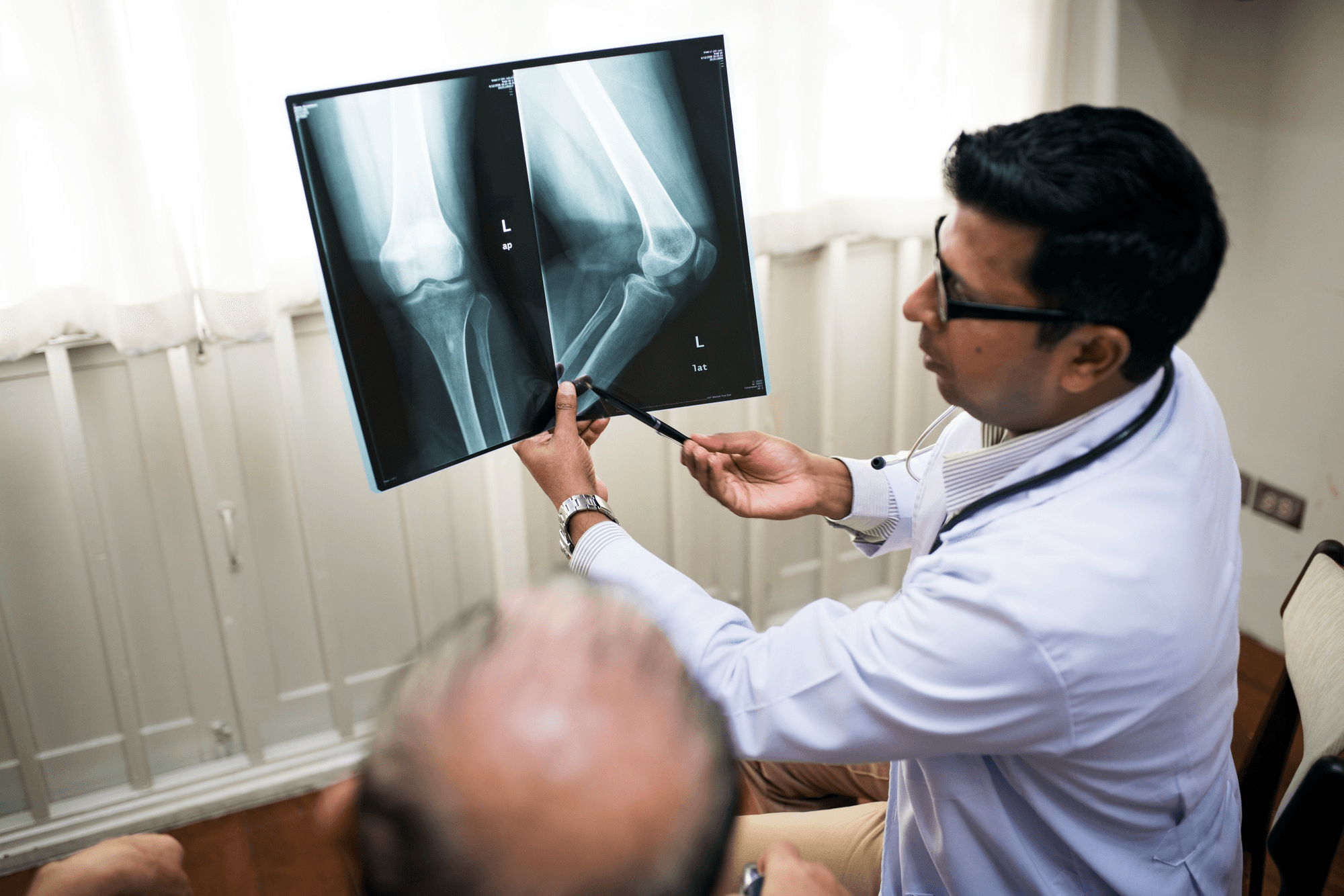
Dr. Mayank Chauhan performs arthroscopic meniscal surgery — both repair and partial meniscectomy — at Prakash Hospital, Sector 33, Noida. Pre-operative MRI evaluation determines tear pattern, zone, and repair feasibility before surgical planning.
To book a consultation, call the number listed on the website.