Rotator Cuff Repair Surgery — How It's Done and What Recovery Looks Like
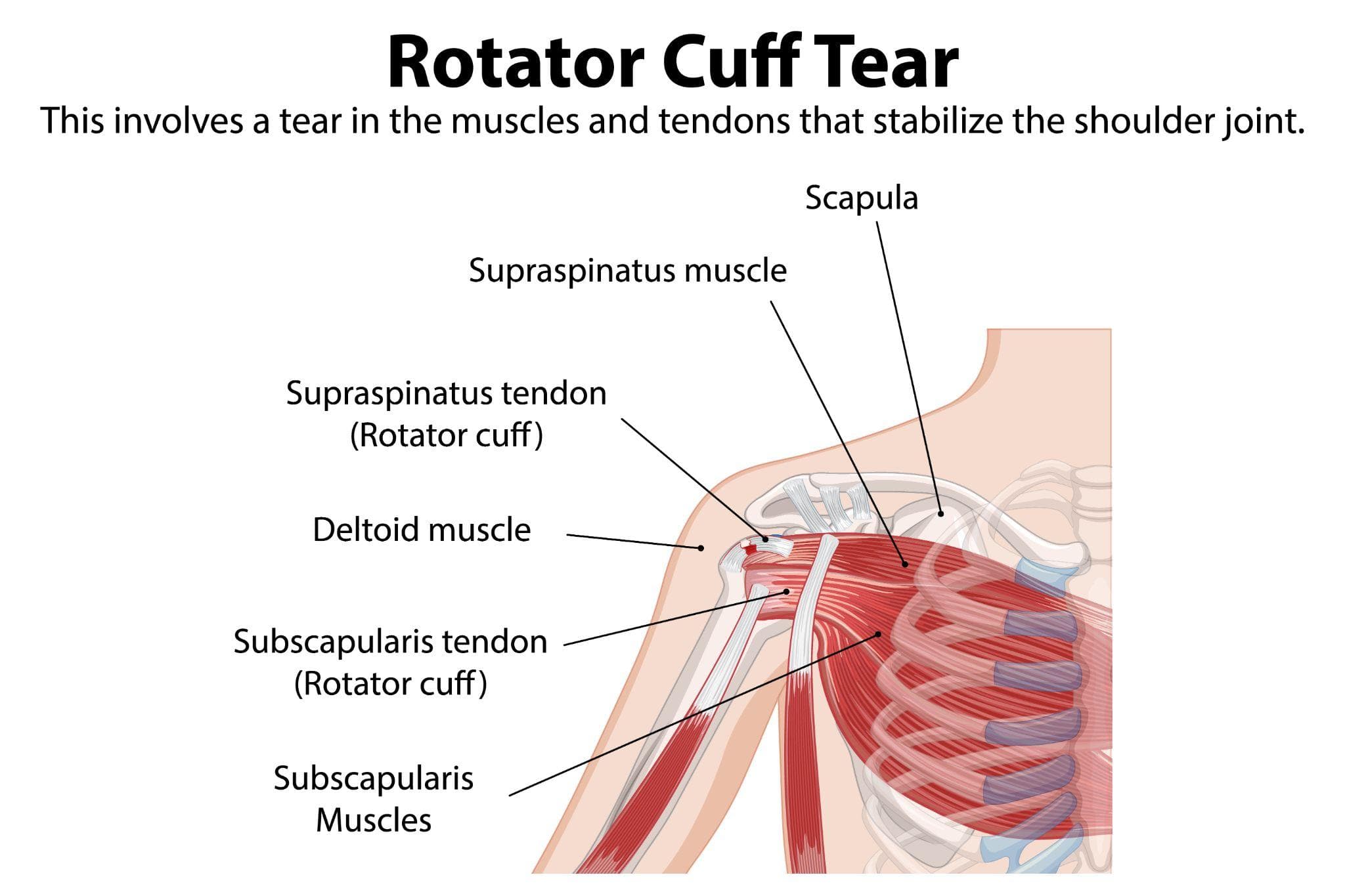
Medical illustration of a rotator cuff tear showing shoulder anatomy with labeled muscles and tendons.
The rotator cuff is one of the most frequently injured structures in the human shoulder — and one that most people have only a vague understanding of until it tears.
The name creates confusion. It's not a single tendon or a protective covering. It's a group of four muscles and their tendons that wrap around the shoulder joint, keeping the humeral head (ball) centred in the glenoid (socket) and providing the power for lifting, rotating, and controlling the arm in all directions.
When these tendons tear — from a fall, from a throwing injury, from years of repetitive overhead activity, or simply from the gradual degeneration that comes with age — the consequences are significant: persistent pain, weakness when lifting, inability to sleep on the affected side, and a shoulder that cannot perform reliably in the positions life demands.
This guide explains when surgery is actually needed, how the repair is performed, and what the recovery realistically looks like.
The Four Muscles and Their Vulnerabilities
Supraspinatus: Runs across the top of the shoulder, attaching to the top of the humeral head. Responsible for initiating arm elevation. The most commonly torn tendon — it passes through a narrow space (the subacromial space) beneath the acromion bone, where impingement and wear are most likely.
Infraspinatus: At the back of the shoulder, responsible for external rotation. Often torn alongside supraspinatus in larger tears.
Subscapularis: At the front of the shoulder, responsible for internal rotation. When this tendon is involved, the functional impact on the arm is significant.
Teres minor: The smallest, rarely torn in isolation.
Most rotator cuff tears involve the supraspinatus, often extending to infraspinatus in larger or more chronic tears. Massive tears involve three or more tendons and represent some of the most surgically challenging shoulder problems.
Does Every Rotator Cuff Tear Need Surgery?
No. And this is worth being clear about upfront.
Tears are classified as:
- Partial thickness tears: Only part of the tendon is torn, the other side remains intact
- Full thickness tears: The tendon is completely torn through, though the torn ends may remain in reasonable proximity
Small partial-thickness tears in older, less active patients frequently respond to physiotherapy, anti-inflammatory medication, and subacromial corticosteroid injection without ever requiring surgery. The body accommodates, other muscles compensate, and function is acceptable.
Surgery is more likely appropriate when:
- The tear is complete (full thickness) and causing significant pain and weakness
- The tear is large or massive
- The patient is younger and physically active — the functional demand justifies repair
- Conservative management has failed after 3–6 months of genuine effort
- The tear occurred from acute trauma (a fall or sudden force) — acute tears have better healing potential after repair than chronic, degenerative tears
- There is progressive weakness
The decision is individualised. Age, activity level, the size and pattern of the tear, and the quality of the remaining tendon tissue all influence whether repair is likely to produce a meaningful functional benefit.
The Operation: Arthroscopic Rotator Cuff Repair
Nearly all rotator cuff repairs performed in India today are done arthroscopically — through small portals using a camera and specialised instruments, without opening the shoulder. This has replaced traditional open repair for the vast majority of cases, with equivalent or superior clinical outcomes and significantly less approach-related trauma.
Anaesthesia
Typically general anaesthesia combined with an interscalene nerve block (numbing the shoulder and arm via the brachial plexus). The nerve block provides excellent pain control for 12–18 hours after surgery, significantly reducing early opioid requirements.
Positioning
Beach chair position (patient sitting at approximately 60 degrees) or lateral decubitus (lying on the opposite side). The surgeon works around the shoulder joint through small portals.
Diagnostic Survey
The arthroscope enters the joint first. The surgeon systematically inspects the shoulder — the rotator cuff from inside the joint, then from the subacromial space above — confirming the tear pattern, assessing the tendon quality and mobility, and looking for any concurrent pathology (biceps tendon involvement, acromial bone spur, labral damage).
Acromioplasty (if indicated)
If a bone spur on the underside of the acromion is present and contributing to impingement, it is smoothed with an arthroscopic burr. This step is not universally necessary and its value in isolated rotator cuff repair is debated.
Preparing the Footprint
The bone surface where the tendon attaches (the greater tuberosity) is prepared with a burr or shaver to create a bleeding bone bed. This vascularised surface improves healing of the repaired tendon.
Suture Anchors
The tendon is re-attached to bone using suture anchors — small metallic or bioabsorbable devices, typically 5–6 mm in diameter, threaded with sutures and screwed into the bone of the greater tuberosity.
Sutures from each anchor are passed through the tendon and tied down, pulling the torn tendon back to its anatomical footprint on the bone. Multiple anchors are typically used depending on tear size — a small tear may need 1–2 anchors, a massive tear may require 4–6 in a double-row configuration.
The double-row technique — placing one row of anchors at the inner edge and a second row at the outer edge of the footprint — creates a broader contact area between tendon and bone and has been shown in multiple studies to produce better footprint coverage and potentially better healing rates than single-row repair.
Wound Closure
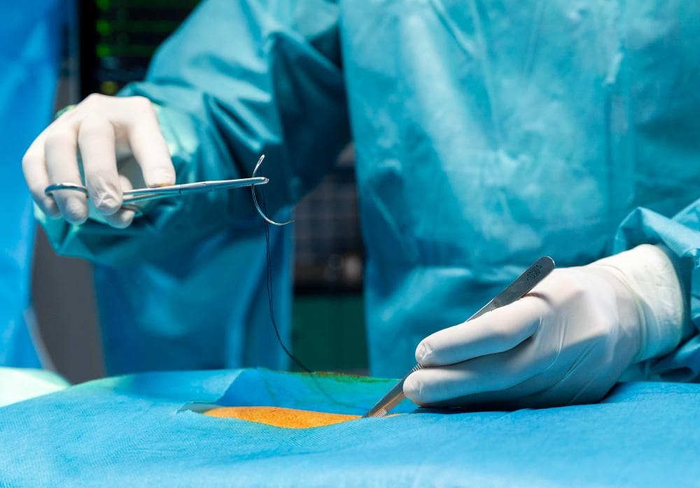
Portals are closed with a suture or steri-strip. A sling is applied. The patient goes to recovery.
Total operative time: 60–120 minutes depending on tear complexity.
Recovery: The Honest Timeline
Rotator cuff repair has one of the longest recovery timelines in elective orthopedic surgery. This is not a reflection of surgical failure — it reflects the biology of tendon-to-bone healing, which is inherently slow.
Weeks 1–6 (Immobilisation Phase): The arm is in a sling for 4–6 weeks. The repair is at its most fragile during this period. Passive range-of-motion exercises (moving the arm with the opposite hand or with a therapist's assistance) begin early to prevent stiffness — but active use of the repaired shoulder is not permitted.
Weeks 6–12 (Active Motion Phase): The sling is removed. Active-assisted exercises begin — the patient starts recruiting the repaired muscles, progressing from gravity-eliminated positions to gravity-resisted movement.
3–6 Months (Strengthening Phase): Progressive resistance training of the rotator cuff and periscapular muscles. This is when most of the functional gains occur.
6–12 Months (Return to Activity): Return to full work, sport, and overhead activity. Most surgeons require 9–12 months before unrestricted overhead sport (cricket bowling, racquet sports, swimming) is permitted.
Desk work: Return possible at 2–6 weeks depending on shoulder requirements.
Physical or overhead work: 4–6 months at minimum.
What Affects the Outcome
Tear size: Small tears repaired early have the best outcomes — 85–90% of patients achieve full pain relief and function. Massive, chronic tears have more variable results.
Tendon quality: Chronically torn tendons become retracted (pulled away from the bone) and fatty-infiltrated (muscle replaced by fat). Severely degenerated tendons may not hold sutures reliably. This is why delaying repair of large tears is counterproductive.
Patient age: Younger patients heal better and have higher functional expectations. Older patients (over 70) with smaller tears and lower activity demands can do well without surgery.
Compliance with rehabilitation: This is the variable most within the patient's control. Patients who follow the rehabilitation programme precisely — not moving the arm actively before six weeks, not missing physiotherapy, performing home exercises consistently — have measurably better outcomes than those who don't.
Shoulder Surgery at Prakash Hospital, Noida
Dr. Mayank Chauhan performs arthroscopic rotator cuff repair at Prakash Hospital, Sector 33, Noida, for patients with symptomatic rotator cuff tears unresponsive to conservative management. Each patient is evaluated with MRI before surgery to assess tear size, pattern, and tendon quality.
To book a consultation, call the number listed on the website.