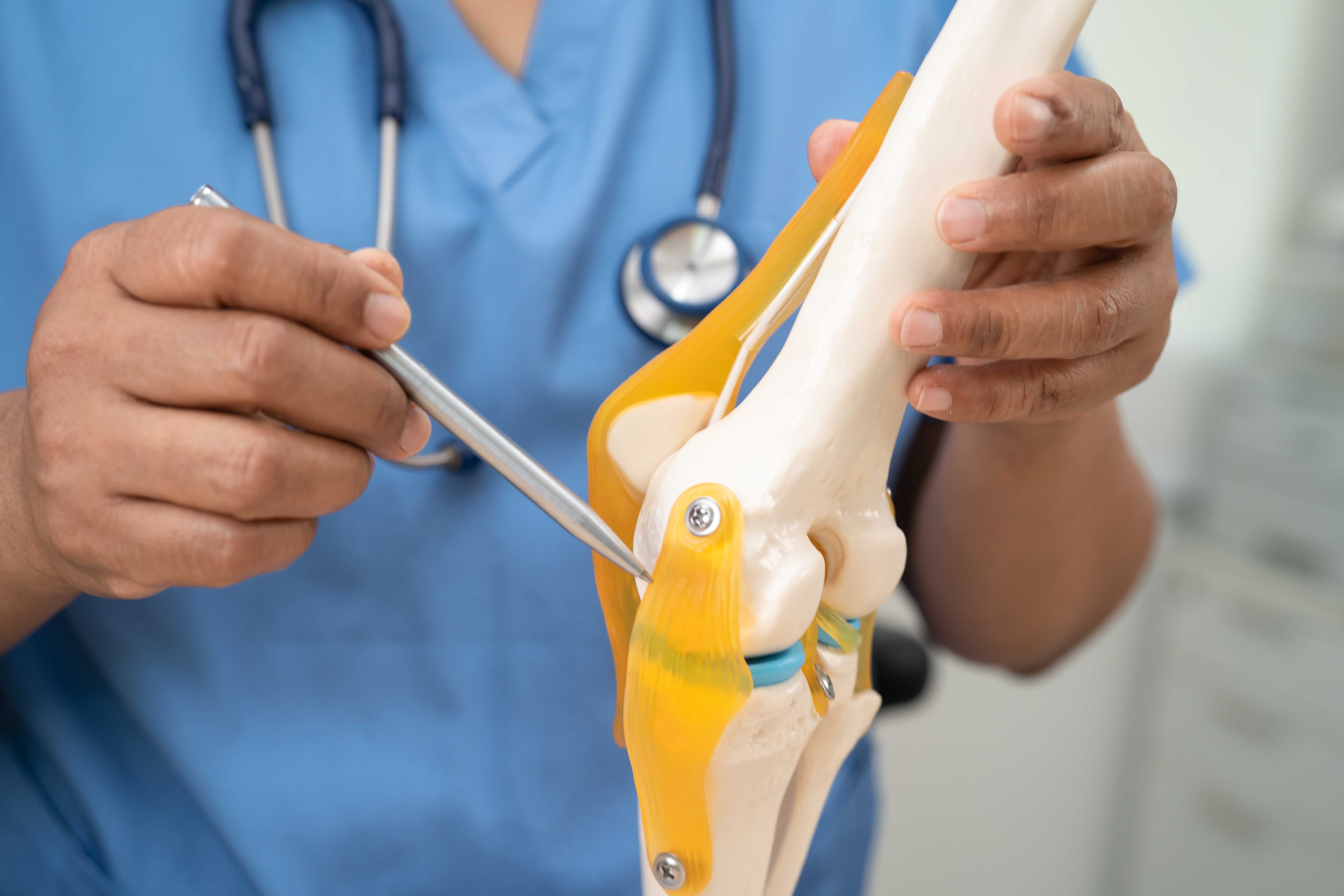
Osteoporosis and Arthritis - Understanding the Link and Managing Both Together

An anatomical illustration comparing a normal bone structure to one affected by severe osteoporosis. On the left, the normal bone displays a dense and healthy structure, while on the right, the severe osteoporosis shows a porous and weakened bone.
In orthopedic clinics across Noida, two conditions are encountered together more often than separately: osteoporosis and arthritis. A 68-year-old woman presenting with knee pain from osteoarthritis almost certainly also has some degree of reduced bone density. An elderly man recovering from a hip fracture — the classic consequence of osteoporosis — frequently has arthritis in the same or other joints.
Despite frequently coexisting, osteoporosis and arthritis are fundamentally different diseases — different in their biology, different in their X-ray appearances, different in their treatment, and different in their implications for surgical management. Understanding this distinction, while recognising how they interact, is essential for patients managing both conditions.
The Fundamental Difference — Two Different Bone Problems
Arthritis (Osteoarthritis):
A disease of the articular cartilage — the smooth, specialised tissue covering the ends of bones within joints. Osteoarthritis is characterised by progressive cartilage breakdown, secondary bone changes (osteophyte formation, subchondral sclerosis), and inflammation of the synovial lining. The bones themselves in OA are typically denser than normal in the affected areas — the subchondral bone (bone immediately below the cartilage) thickens and hardens as a response to increased mechanical stress.
Osteoporosis:
A disease of bone density and quality — affecting the entire skeleton, not specific joints. In osteoporosis, the trabecular (spongy) architecture within bones becomes thinner, the cortical (outer) bone shell becomes thinner, and the overall bone mineral density falls below the threshold that can safely withstand normal loads. The joints themselves may be completely normal in early osteoporosis — it is the bone between and below joints that is affected, not the articular surface.
The paradox: In some ways, these conditions are opposites in their bone effects. OA increases bone density at the joint surface (subchondral sclerosis, osteophytes — dense bone). Osteoporosis decreases bone density throughout the rest of the skeleton. A patient can have both simultaneously — dense bone at the arthritic knee joint surface and thin, fragile bone in the spine and hip.
Why They Frequently Coexist
Despite being different diseases, osteoporosis and arthritis share demographic risk factors that cause them to occur together:
Age: Both increase dramatically with age. Women over 65 are at high risk of both conditions.
Post-menopausal hormonal change: The loss of oestrogen at menopause simultaneously increases osteoclastic bone resorption (driving osteoporosis) and reduces the anti-inflammatory protection that oestrogen provided to joints. Postmenopausal women are therefore at increased risk of both.
Low physical activity: Sedentary behaviour weakens bones (less mechanical load stimulus for bone formation) and weakens the muscles that protect joints. Inactivity contributes to both conditions.
Obesity: Overweight individuals have higher mechanical loads on weight-bearing joints (driving OA) while also having altered hormonal and metabolic profiles that can affect bone metabolism. In South Asian populations specifically, the combination of central obesity with Vitamin D deficiency creates a pattern of joint loading combined with poor bone quality.
Vitamin D deficiency: Drives bone demineralisation (osteoporosis) and worsens arthritis through its pro-inflammatory effects (as described in the Vitamin D blog).
Rheumatoid Arthritis and Osteoporosis — A Special Relationship
The relationship between osteoporosis and rheumatoid arthritis (RA) is even stronger than with osteoarthritis.
RA causes osteoporosis through multiple mechanisms:
Systemic inflammation: The chronically elevated levels of inflammatory cytokines (particularly IL-6, TNF-α) in RA stimulate osteoclast activity — the bone-resorbing cells. This produces systemic bone loss throughout the skeleton, not just at the inflamed joints.
Periarticular bone loss: Around inflamed RA joints, the intense local inflammatory environment causes accelerated periarticular osteopenia — visible as juxtaarticular (near-joint) rarefaction on X-ray. The "periarticular osteopenia" sign is actually a diagnostic feature of RA on X-ray.
Corticosteroid treatment: Many RA patients receive prolonged courses of oral corticosteroids (prednisolone). Long-term steroid use is the most important drug-related cause of osteoporosis — inhibiting osteoblast function and increasing urinary calcium loss. Any RA patient on long-term steroids should be on bone-protective treatment (bisphosphonate) unless contraindicated.
Reduced mobility: RA-related joint disease limits physical activity — reducing the mechanical load that drives bone formation.
The clinical implication: All patients with rheumatoid arthritis should have bone density assessed (DEXA scan) — particularly those on corticosteroids — and should receive bone-protective treatment as appropriate.
How to Distinguish Osteoporosis from Arthritis on Investigation
X-Ray Differences
| Feature | Osteoarthritis | Osteoporosis |
|---|---|---|
| Joint space | Narrowed | Normal (until fragility fracture occurs) |
| Bone density at joints | Increased (subchondral sclerosis) | Reduced throughout |
| Osteophytes | Present | Absent |
| Fractures | Absent (unless combined) | Present (vertebral compression fractures, hip fractures) |
| Global bone appearance | Normal or dense | Radiolucent (bones appear darker — less dense) |
DEXA Scan
The definitive investigation for osteoporosis measures bone mineral density at the lumbar spine and hip, reported as T-scores. Does not diagnose arthritis.
Blood Tests
Arthritis: X-ray and clinical assessment. RA requires specific tests (RF, anti-CCP, inflammatory markers).
Osteoporosis: Calcium, Vitamin D, PTH, renal function, bone turnover markers. Rule out secondary causes (hyperparathyroidism, coeliac disease, multiple myeloma).
The Interaction — How Each Makes the Other Worse
The most clinically important interaction is how osteoporosis affects the management of arthritis — particularly surgical management.
Osteoporosis and Knee Replacement
In a patient with both knee OA (needing replacement) and significant osteoporosis, several specific concerns arise:
Implant fixation: Total knee replacement implants are fixed to the femur and tibia — either with bone cement or by press-fit (cementless) fixation relying on bone ingrowth. In osteoporotic bone, press-fit cementless fixation is less reliable — the porous bone may not provide adequate initial stability or long-term ingrowth. Cemented fixation is typically preferred in patients with low bone density.
Periprosthetic fracture risk: Osteoporotic bone around the implant is more vulnerable to fracture — either at the time of surgery (intraoperative) or subsequently from relatively minor trauma. These fractures are complex to manage and have worse outcomes than in normal bone.
Tibial component sinkage: The tibial component of a knee replacement is supported by the tibial plateau — the flat top surface of the tibia. Osteoporotic tibial bone provides a weaker foundation, with a higher risk of the component gradually subsiding into the soft bone over the years.
What this means practically:
- Bone density should be assessed before major joint replacement surgery in elderly patients or those at risk of osteoporosis
- If significant osteoporosis is found, treatment (bisphosphonate) should ideally be started before elective replacement to improve bone quality
- Surgical technique and implant choice should be adapted for the bone quality present
Osteoporosis and Hip Fractures
The most dramatic interaction: a fall in a patient with osteoporosis + hip arthritis (or AVN) produces a hip fracture at a site that is already compromised.
Hip fractures — femoral neck and intertrochanteric fractures — are almost exclusively a consequence of osteoporosis in the elderly. The pathological sequence: reduced bone density → fall → fracture → surgical repair or hemiarthroplasty → recovery complicated by arthritis, deconditioning, and medications associated with both underlying conditions.
Preventing the first fracture — through DEXA screening, Vitamin D and calcium supplementation, bisphosphonate therapy, and fall prevention — is infinitely preferable to treating it after it occurs.
Managing Both Conditions Simultaneously
Many patients in Noida's older population have both OA and osteoporosis — requiring management of both simultaneously. The key principles:
1. Don't Let OA Treatment Worsen Osteoporosis
Some treatments used for arthritis pain are problematic for bone health:
- Corticosteroids (oral prednisolone): Regular use for arthritis pain significantly worsens bone density. Use the minimum necessary dose for the shortest necessary period. If prolonged steroid use is unavoidable, prescribe bone protection (bisphosphonate + calcium + Vitamin D) simultaneously.
- Excessive bed rest: Inactivity for arthritis pain reduces bone loading and accelerates bone loss. Appropriate weight-bearing exercise — even if modified for joint pain — maintains bone density.
2. Exercise for Both Conditions
Weight-bearing exercise addresses both OA and osteoporosis simultaneously:
- Walking: Maintains bone density at the hip and spine AND strengthens the muscles that protect arthritic joints AND reduces body weight (reducing joint load)
- Resistance training: Stimulates bone formation AND strengthens muscles that support arthritic joints
- Aquatic exercise: For patients with severe OA who cannot tolerate weight-bearing exercise — maintains muscle strength and cardiovascular fitness without joint loading, though with less bone-protective stimulus
The key is finding exercises the patient can do given their joint limitations — the right physiotherapist can design a programme that works around the arthritis while providing the loading stimulus needed for bone maintenance.
3. Nutrition — Addressing Both Simultaneously
Calcium (1200 mg daily), Vitamin D (supplements to achieve a serum level above 30 ng/mL), and adequate protein benefit both conditions. Anti-inflammatory dietary choices benefit arthritis. There is no dietary conflict between managing osteoporosis and arthritis simultaneously.
4. Medication Coordination
Patients on bisphosphonates (for osteoporosis) who need joint surgery require specific planning — bisphosphonates are associated with impaired fracture healing and osteonecrosis of the jaw in rare cases. The timing of bisphosphonate treatment relative to elective surgery should be discussed with the prescribing physician and operating surgeon.
Patients on NSAIDs for arthritis should be aware that very long-term NSAID use has been associated with modest effects on bone metabolism in some studies — though this is not a sufficient reason to avoid NSAIDs when clinically needed.
Osteoporosis and Arthritis Management in Noida — Dr. Mayank Chauhan at Prakash Hospital
Dr. Mayank Chauhan, Senior Orthopedic Surgeon at Prakash Hospital, Sector 33, Noida, evaluates and manages both conditions in the context of the whole patient — ensuring that the evaluation of a patient presenting with knee or hip pain includes an assessment of bone density where indicated, and that any joint replacement surgery considers and accounts for the bone quality present.
For elderly patients in Noida and Greater Noida with both joint pain and a history of fractures or known osteoporosis, a comprehensive orthopedic consultation considers both elements of their bone health.
To book a consultation, call the number listed on the website.
The Bottom Line
Osteoporosis and arthritis frequently coexist but are fundamentally different conditions — affecting different aspects of bone and joint health, with different implications and different treatments. Understanding both, addressing both, and ensuring they are managed in coordination — particularly in patients facing joint surgery — produces better outcomes than treating each in isolation.
The shared preventive strategies — weight-bearing exercise, calcium, Vitamin D, fall prevention — serve both conditions simultaneously. Use them.
To consult Dr. Mayank Chauhan, Senior Orthopedic Surgeon in Noida, call the number listed on the website.