Hip Bursitis — Causes, Symptoms, and Treatment Without Surgery
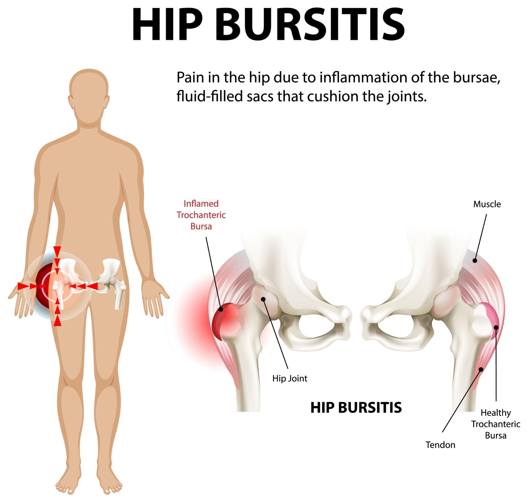
A diagram explaining hip bursitis. The body shows the location of hip bursitis and a highlighted red region where pain occurs. A diagram also shows the human hip joint with labels and highlights inflammation in the trochanteric bursa.
Hip pain that worsens when lying on your side, hurts most on the outer part of the hip, and gets worse with stairs or prolonged sitting is not always arthritis. In many cases — particularly in middle-aged to older women — it is hip bursitis, specifically trochanteric bursitis.
It's a diagnosis that is frequently missed or misattributed. Patients are told they have "hip arthritis" without a proper examination, given vague activity restriction advice, and sent home still in pain.
Bursitis is distinctly different from arthritis, responds to different treatment, and in the majority of cases resolves completely without surgery.
What Is a Bursa?
A bursa (plural: bursae) is a small, fluid-filled sac that acts as a cushion between structures that would otherwise rub against each other — tendons against bone, or bone against skin. The body has over 150 bursae. Most are tiny and you're never aware of them. When they become inflamed — bursitis — they swell, become tender, and cause pain with movement.
Around the hip, there are several bursae. The two clinically significant ones are:
The trochanteric bursa: Located over the greater trochanter — the prominent bony bump you can feel on the outer side of the hip. The iliotibial band (IT band) and the gluteal tendons pass over this bony prominence, and the bursa cushions this interface. Inflammation here is called greater trochanteric bursitis or trochanteric pain syndrome.
The iliopsoas (iliopectineal) bursa: Located at the front of the hip, between the iliopsoas muscle and the hip joint. Iliopsoas bursitis causes groin pain and is less common than trochanteric bursitis.
What Causes Hip Bursitis?
Bursitis develops when the bursa is irritated — usually by repetitive friction, abnormal biomechanics, or direct trauma.
Repetitive friction: Activities that involve repeated movement of the IT band or gluteal tendons over the greater trochanter — running, cycling, stair climbing, walking long distances — can inflame the bursa over time.
Muscle weakness: Weakness in the hip abductor muscles (gluteus medius and minimus) is a major contributing factor to trochanteric bursitis. When these muscles are weak, the pelvis tilts excessively during walking (a pattern called Trendelenburg gait), increasing friction at the greater trochanter.
Leg length discrepancy: Even a minor difference in leg length (1–2 cm) can alter the way the hip moves and increase IT band tension on the affected side.
Hip surgery: Prior hip replacement surgery can occasionally cause trochanteric bursitis if the hardware or soft tissue reconstruction affects the mechanics of the tendons over the greater trochanter.
Direct trauma: A fall directly onto the outer hip can inflame the bursa.
Inflammatory conditions: Rheumatoid arthritis and other inflammatory conditions can cause bursitis as part of a broader joint inflammation.
Age and sex: Trochanteric bursitis is more common in middle-aged to older women. Hormonal factors and the wider female pelvis (which increases the angle of the IT band) are likely contributors.
Symptoms: What Does Hip Bursitis Feel Like?
Trochanteric bursitis has a fairly characteristic presentation that distinguishes it from hip joint arthritis:
Location of pain: The outer side of the hip and thigh. Not in the groin (which is more typical of true hip joint pathology). Not in the buttock (which suggests sciatic or piriformis involvement). The outer hip.
Tenderness: Direct pressure over the greater trochanter — you can press on it and reproduce the pain. This is a simple bedside test that points strongly toward bursitis rather than joint arthritis.
Lying on the affected side: Typically very painful. Many patients wake at night because they have rolled onto the affected side.
Stairs: Walking upstairs (which loads the abductor muscles and IT band) typically worsens the pain.
Prolonged walking or standing: Also aggravating. Rest relieves pain initially, but extreme rest without rehabilitation makes the underlying muscle weakness worse.
Hip range of motion: Usually preserved. Unlike hip joint arthritis — where internal rotation in particular is restricted and painful — trochanteric bursitis does not characteristically limit the range of hip joint movement. This distinction is helpful on examination.
How Is It Diagnosed?
The diagnosis is primarily clinical — based on history and physical examination. Key examination findings:
- Tenderness on palpation of the greater trochanter
- Pain reproduced with resisted hip abduction (the patient pushes their leg outward against resistance)
- Normal or near-normal hip joint range of motion
- Possible Trendelenburg sign (the opposite hip drops when standing on the affected leg)
X-rays of the hip are typically normal or show only the underlying joint, which helps rule out hip joint arthritis as the primary diagnosis.
MRI can visualise bursal swelling and — importantly — any associated tendon pathology (gluteus medius or minimus tears, which can coexist with bursitis and significantly affect treatment planning).
Ultrasound is useful for confirming bursal swelling and guiding injections.
Treatment: Most Cases Resolve Without Surgery
Physiotherapy
The most important component of treatment — and the one most often skipped.
Hip bursitis is frequently driven by gluteal muscle weakness. The bursa is inflamed because the hip abductor mechanism is not functioning efficiently, putting excess stress on the tissue overlying the greater trochanter. Treating only the inflammation without addressing the underlying weakness leads to recurrence.
A physiotherapist-directed programme focuses on:
- Gluteus medius strengthening: Side-lying hip abduction, clamshells, single-leg exercises
- IT band flexibility: Specific stretching to reduce lateral hip tension
- Hip flexor and hamstring stretching: Poor flexibility in these groups affects gait mechanics
- Gait retraining: Addressing Trendelenburg gait pattern and excessive pelvic drop
Results from physiotherapy are not immediate. A meaningful programme takes 6–12 weeks. Patients who abandon physiotherapy after 2 weeks are making a mistake.
Activity Modification
Reducing the activities that most aggravate the bursa — typically high-impact running, excessive stair climbing, or prolonged walking — while maintaining general conditioning with swimming or cycling (which do not stress the outer hip).
Avoid sleeping on the affected side. Placing a pillow between the knees when sleeping on the opposite side reduces lateral hip tension.
Anti-Inflammatory Medication
NSAIDs (ibuprofen, naproxen, diclofenac) can reduce bursal inflammation and provide pain relief. Most useful for short-term relief while physiotherapy works on the underlying cause. Not a long-term solution.
Corticosteroid Injection
For patients with significant pain that is limiting physiotherapy, a corticosteroid injection directly into the bursa — typically performed under ultrasound guidance — can dramatically reduce inflammation within days, providing a window for physiotherapy to progress.
Injections are effective for short-to-medium term relief. They are not a cure — the underlying biomechanical issues must still be addressed to prevent recurrence. More than 2–3 injections at the same site over 12 months is generally not recommended.
PRP Injection
For patients with recurrent bursitis or associated tendon degeneration, platelet-rich plasma (PRP) injection is an emerging option. The evidence for PRP in greater trochanteric bursitis is growing, particularly when there is associated gluteal tendinopathy.
When Is Surgery Considered?
Surgery is rarely required and is considered only after a genuine 3–6 month trial of conservative management has failed.
Surgical options include:
- Arthroscopic bursectomy: Removal of the inflamed bursa through small portals. Effective for isolated bursitis, with good outcomes in carefully selected patients.
- IT band release: In cases where IT band tightness over the trochanter is a primary driver, a small release of the band can reduce friction.
- Gluteal tendon repair: If a full-thickness gluteal tendon tear is identified on MRI as a contributing factor, surgical repair may be required.
These are uncommon interventions. The vast majority of trochanteric bursitis cases resolve with physiotherapy and, when needed, injection.
Hip Pain Assessment in Noida
Hip pain on the outer side of the hip is often bursitis — but distinguishing it from hip joint arthritis, referred lumbar pain, or other sources requires a proper examination and appropriate imaging.
Dr. Mayank Chauhan at Prakash Hospital, Sector 33, Noida, evaluates hip pain comprehensively, distinguishing between conditions that require surgery and those that respond to conservative management. Not every hip that hurts needs replacing.
To book a consultation, call the number listed on the website.