Stress Fractures - What They Are, Why They Happen, and How to Treat Them
person with an avulsion fracture
Stress fractures are uniquely deceptive injuries. They don't happen from a single traumatic event — no fall, no collision, no obvious moment of injury. They develop silently, over weeks, as a bone subjected to repetitive loading begins to fail at the microscopic level. By the time pain is significant enough to limit activity, the bone has already accumulated substantial structural damage.
In Noida's growing community of runners, cricketers, fitness enthusiasts, military recruits, and young athletes, stress fractures are increasingly common — and increasingly the subject of delayed diagnosis. The combination of gradual onset, normal early X-rays, and the cultural tendency among athletes to "train through pain" creates conditions where a stress fracture can progress from a bone stress reaction to a complete fracture before anyone takes it seriously.
The Biology of a Stress Fracture
Bone is not a static structure. It is continuously remodelling — breaking down old bone (osteoclastic resorption) and building new bone (osteoblastic formation). This remodelling process is the mechanism by which bone adapts to changing loads.
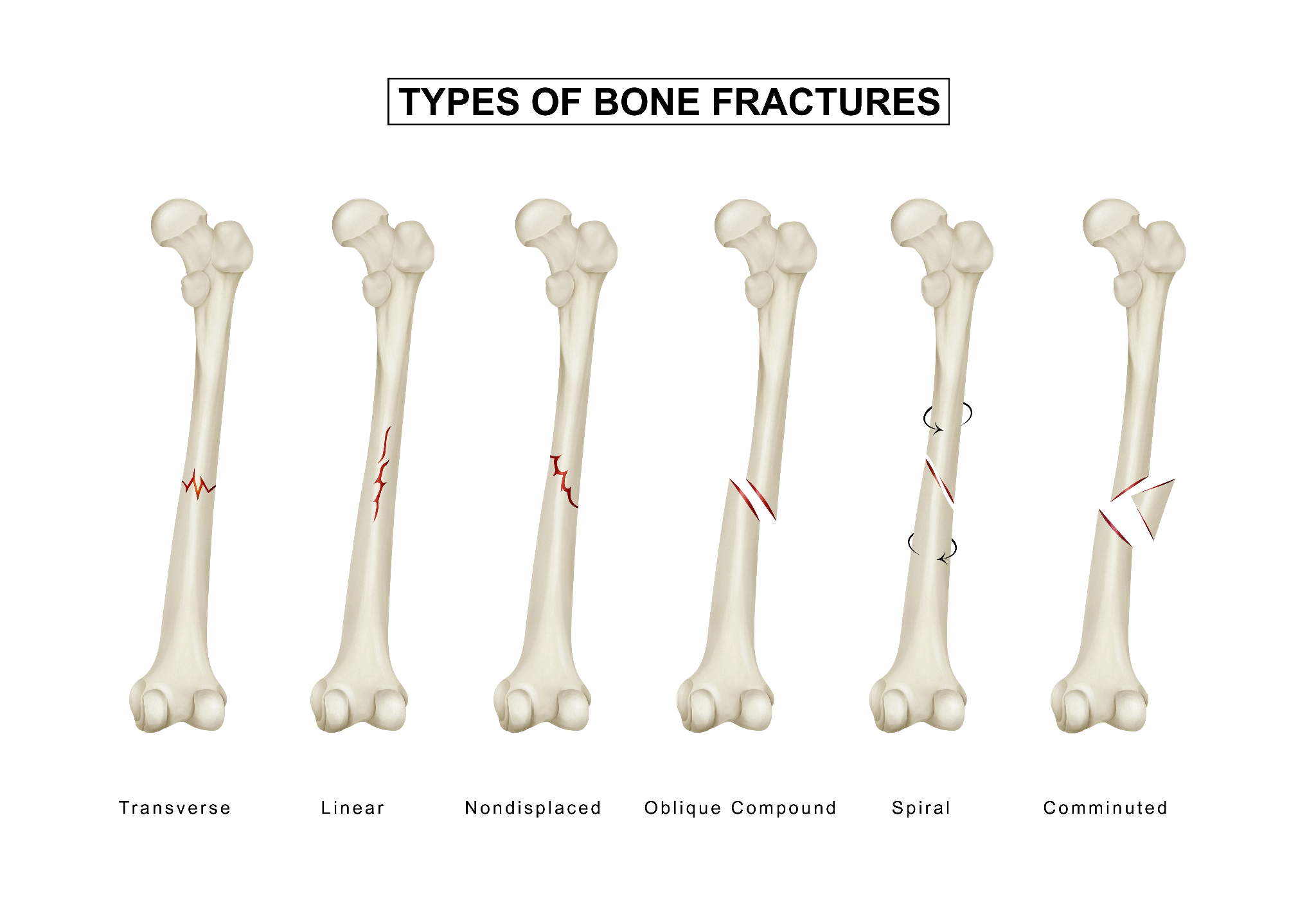
When loading exceeds the bone's capacity to remodel adequately, microdamage accumulates faster than it can be repaired. This progressive accumulation of microdamage is a bone stress reaction — a continuum that, if loading continues, progresses to a stress fracture (a visible crack in the bone cortex), and ultimately to a complete fracture (the bone breaks through entirely).
The critical determinant is the balance between:
- Loading rate: How frequently and intensely the bone is being stressed
- Remodelling capacity: How effectively the bone can repair microdamage between loading sessions
When training load increases faster than the bone's capacity to adapt — as happens with sudden increases in running mileage, a new training programme, or military basic training — stress fractures occur.
Who Gets Stress Fractures?
Athletes — Overuse and Training Errors
Runners: Tibial (shin bone) and metatarsal (foot bone) stress fractures are the classic runner's injuries. Sudden increases in weekly mileage, switching from soft to hard surfaces, or inadequate footwear are the most common triggering factors.
Military recruits: Basic training creates the perfect conditions for stress fractures — high-intensity loading in unfit individuals with insufficient rest. Tibia and metatarsal fractures are most common.
Cricket fast bowlers: Lumbar pars interarticularis stress fractures are specific to fast bowling — the extreme lumbar extension-rotation of the bowling action creates cyclical stress on this structure. The most common career-limiting injury in young Indian pacers. Covered in the cricket injuries blog.
Dancers and gymnasts: Metatarsal (foot), tibia, and fibula stress fractures from the repetitive impact and landing loads.
Badminton and basketball players: Tibial and fibular stress fractures from the repeated jumping and pivoting.
Football players: Metatarsal stress fractures — the "March fracture" or fifth metatarsal stress fracture — are among the most common injuries in elite football. The classic mechanism of stopping suddenly while running stresses the fifth metatarsal.
Non-Athletes — Bone Quality Issues
Stress fractures are not exclusive to athletes. Anyone whose bones are subjected to repetitive loading beyond their structural capacity is at risk:
Osteoporotic patients: Insufficiency fractures (a form of stress fracture) occur in bone that is too weak to handle normal daily loading. Metatarsal insufficiency fractures are common in elderly women with osteoporosis. Sacral insufficiency fractures cause low back and buttock pain without trauma.
Vitamin D and calcium deficiency: Inadequate mineralisation reduces bone quality and increases stress fracture risk — particularly relevant in Noida's indoor-working, sun-avoiding, dairy-insufficient population.
The Female Athlete Triad: The combination of low energy availability (often from disordered eating), low bone mineral density, and menstrual dysfunction creates dramatically increased stress fracture risk in female athletes — and this triad is increasingly recognised in Indian female athletes and dancers.
Common Stress Fracture Locations — and Why Each One Matters
Tibia (Shin Bone)
The most common stress fracture in runners and military recruits. Pain is felt along the inner border of the tibia — typically in the middle or lower third of the shin.
Medial tibial stress syndrome (shin splints) vs. tibial stress fracture:
This is a critical distinction. Shin splints (medial tibial stress syndrome) produce pain along a longer segment of the tibia's inner border, diffuse on palpation, related to compartment pressure and periosteal stress. A tibial stress fracture produces point tenderness — pressing on a specific small area reproduces the exact pain. The distinction requires clinical assessment and MRI.
High-risk tibial fracture (anterior tibial stress fracture):
The anterior cortex of the tibia (front shin surface) carries tensile forces during running — the bone is under tension on the front surface during foot strike. Stress fractures here (producing the "dreaded black line" on MRI — a transverse radiolucent line) are at risk of complete fracture and require aggressive management, including possible surgery (intramedullary nailing) rather than just rest.
Metatarsals (Foot Bones)
The five metatarsals are among the most commonly fractured bones in sports. The second and third metatarsals are most often affected in runners (the classic "March fracture" of soldiers walking long distances). The fifth metatarsal has specific variants:
Fifth metatarsal stress fractures:
- Avulsion fractures at the base: Not true stress fractures — caused by the peroneus brevis tendon pulling off a fragment with ankle inversion.
- Jones fractures: Stress fractures at the junction of the base and shaft of the fifth metatarsal. High non-union rate; often requires surgical fixation with an intramedullary screw for athletes. One of the most important metatarsal fractures to identify correctly because conservative management fails in a significant proportion.
- Shaft fractures: Typically heal with conservative management.
Navicular (Foot)
The navicular is a small bone on the inner midfoot — a relatively uncommon but high-risk stress fracture. Often delayed in diagnosis because pain is vague and the navicular is difficult to assess on plain X-ray (CT or MRI required). Strict non-weight-bearing is typically necessary for 6–8 weeks. Non-union is common without adequate management.
Femoral Neck (Hip)
The femoral neck connects the femoral head (the ball of the hip joint) to the shaft. Stress fractures here are less common but among the most serious.
Compression side (inferior femoral neck): These fractures are on the compression (lower) surface of the femoral neck. They are relatively stable and often heal with rest and protected weight-bearing, though surgical fixation (with cannulated screws) is recommended for athletes.
Tension side (superior femoral neck): Fractures on the tension (upper) surface of the femoral neck are high-risk — they can displace and progress to complete fractures with avascular necrosis. Any femoral neck stress fracture with tension-side involvement should be evaluated immediately by an orthopedic surgeon. Surgical fixation is typically required. Displaced femoral neck fractures in young athletes are devastating, career-ending, and sometimes life-altering injuries.
Who gets femoral neck stress fractures: Runners who have significantly increased mileage, military recruits in infantry training, and patients with osteoporosis. Any athlete with groin or hip pain that worsens with impact activity and is not responding to rest should have an MRI to exclude a femoral neck stress fracture.
Lumbar Spine — Pars Interarticularis (Spondylolysis)
Specific to fast bowlers, gymnasts, and athletes performing repeated lumbar extension and rotation. The pars interarticularis — a small bony bridge at the back of each vertebra — is subjected to shear forces during these activities and can develop a stress fracture (spondylolysis).
Bilateral pars fractures allow the vertebra to slip forward on the one below (spondylolisthesis). Covered in the cricket injuries blog.
Diagnosis
Why X-rays Miss Stress Fractures
Plain X-rays are negative in approximately 70% of stress fractures at initial presentation. The bone changes of a stress fracture — periosteal reaction, endosteal reaction, the fracture line itself — develop slowly and may only become visible on X-ray 2–3 weeks after the fracture begins. Waiting for positive X-ray findings before diagnosing a stress fracture leads to a delayed diagnosis that allows progression.
MRI — The Gold Standard
MRI is the most sensitive imaging investigation for stress fractures. It shows:
- Bone marrow oedema: The earliest change — appears within days of a bone stress reaction beginning
- Periosteal oedema: Swelling around the bone cortex
- The fracture line itself: Visible when a frank crack has developed
- Grade of injury: MRI allows grading of stress injuries from simple stress reaction to complete fracture — guiding the urgency and nature of management
MRI is the investigation of choice whenever a stress fracture is suspected, and X-rays are negative or inconclusive.
Bone Scan
An alternative to MRI — bone scan is highly sensitive for stress fractures (positive within days of injury) but less specific. Can miss the exact location and the fracture line detail. Still useful when MRI is not available.
CT Scan
Particularly useful for:
- Navicular stress fractures (better bony detail than MRI)
- Lumbar pars interarticularis fractures (CT shows the fracture line clearly)
- Assessing fracture healing
Treatment
The General Principle: Reduce Load, Allow Healing
The fundamental treatment for stress fractures is reducing the loading on the bone below the threshold that caused the fracture, while allowing bone remodelling to repair the damage.
Low-risk stress fractures (most metatarsals, fibula, ribs):
- Relative rest — stopping the causative activity but not complete immobilisation
- Walking boot or supportive footwear
- Cross-training (swimming, cycling) to maintain fitness without bone loading
- Return to sport in 6–8 weeks
Moderate-risk stress fractures (tibial shaft, lesser metatarsals):
- Protected weight-bearing with crutches if needed
- Boot or supportive device
- Return to running in 8–12 weeks after serial confirmation of healing
High-risk stress fractures (anterior tibia, navicular, femoral neck tension side, fifth metatarsal Jones fracture):
These require more aggressive management — often surgical fixation for athletes who cannot accept prolonged return-to-sport timelines — due to their high complication rates (non-union, displacement, avascular necrosis) with conservative management.
Surgical Management
Intramedullary nailing for anterior tibial stress fractures: For the "dreaded black line" fracture that risks completion, an IM nail provides stability that allows continued activity and reduces non-union risk.
Screw fixation for navicular and Jones fractures: A single intramedullary screw placed through the fracture significantly improves union rates in athletes who need early return to sport.
Cannulated screw fixation for femoral neck fractures: For all tension-side and many compression-side femoral neck stress fractures — prevents displacement that could lead to avascular necrosis.
Addressing the Underlying Cause
Treatment of the fracture alone is not sufficient. The factors that caused the bone to fail must be addressed:
Training load modification: Return to sport after a stress fracture must be gradual — a 10% per week increase in mileage or training volume is the standard guideline.
Nutritional assessment: Calcium and Vitamin D deficiency must be identified and corrected. Iron deficiency (affecting oxygen delivery to bone) is another common contributor.
Biomechanical assessment: Running gait analysis, footwear assessment, and sports technique review identify mechanical factors that concentrate stress on specific bones.
The Female Athlete Triad assessment: In female athletes, a stress fracture is a sentinel event that should prompt evaluation of energy availability, menstrual status, and bone density.
Stress Fracture Management in Noida — Dr. Mayank Chauhan at Prakash Hospital
Dr. Mayank Chauhan, Senior Orthopedic Surgeon at Prakash Hospital, Sector 33, Noida, evaluates and manages stress fractures across the full spectrum — from early bone stress reactions identified on MRI through to high-risk fractures requiring surgical intervention.
For runners, cricketers, gym-goers, and other athletes in Noida and Greater Noida with activity-related bone pain that persists beyond 2 weeks without clear cause, MRI evaluation for a stress fracture is appropriate. To book a consultation, call the number listed on the website.
The Bottom Line
Stress fractures develop silently, are missed on X-ray in 70% of early cases, and can progress from a manageable bone stress reaction to a complete fracture if activity continues. Early diagnosis with MRI — and immediate load reduction — prevents this progression.
Not all stress fractures are equal. Some heal reliably with 6 weeks of rest. Others — the anterior tibia, navicular, femoral neck, Jones fracture — require specific intervention and careful management to avoid serious complications. When bone pain develops gradually in an athletic context — take it seriously sooner rather than later.
To consult Dr. Mayank Chauhan, Senior Orthopedic Surgeon in Noida, call the number listed on the website.