How ACL Surgery Is Done — A Complete Patient Guide
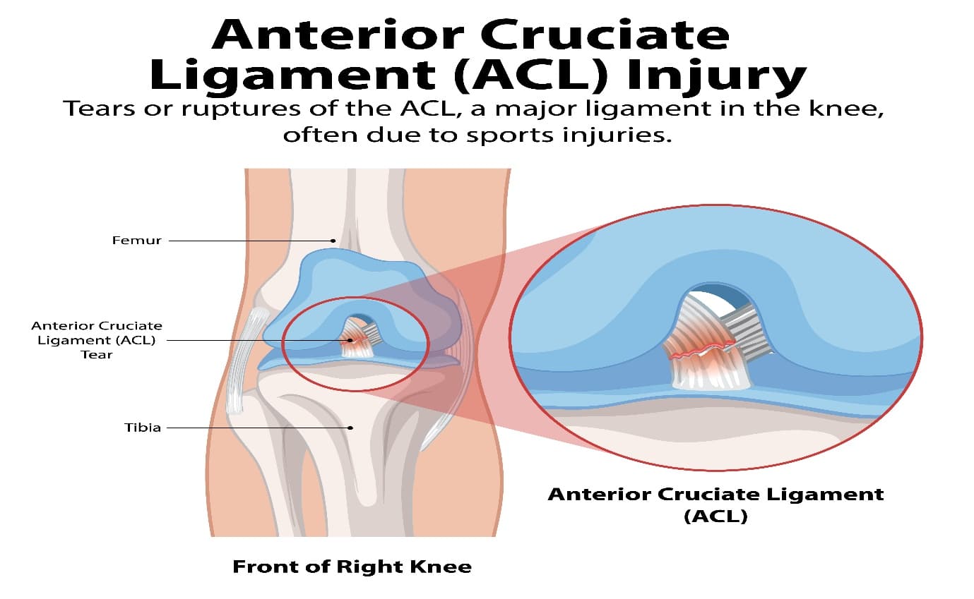
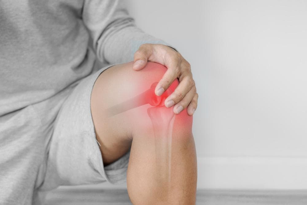
Detailed medical illustration of an anterior cruciate ligament (ACL) injury in the knee.
An ACL tear is one of those diagnoses that lands with a particular kind of dread. You know what it means — weeks in a brace, months of physiotherapy, and the question hanging over everything: will it ever feel the same?
Before any of that conversation can happen productively, it helps to understand what the surgery actually involves. Not the vague "they replace the ligament" summary, but the actual procedure — which graft, which technique, which fixation, and what happens in the months after.
This guide is for patients who have been told they need ACL surgery and want to understand it properly before they walk into the operating room.
What the ACL Does and Why It Tears
The anterior cruciate ligament runs diagonally through the centre of the knee, connecting the femur (thigh bone) to the tibia (shin bone). Its primary role is to prevent the tibia from sliding forward on the femur — the movement that happens every time you pivot, change direction suddenly, or decelerate rapidly.
ACL tears most commonly occur in non-contact situations: landing awkwardly from a jump, planting a foot and rotating, or decelerating suddenly in a sport. The classic mechanism is a planted foot combined with a twisting movement of the body above it. Cricket fielding, football, basketball, kabaddi, and running sports all generate these forces regularly.
Female athletes tear their ACLs at 4–6 times the rate of male athletes with comparable activity levels — a combination of anatomical differences (wider pelvis, greater valgus alignment at the knee), hormonal influences on ligament laxity, and differences in neuromuscular firing patterns.
When the ACL tears, the knee typically gives out immediately. There's often a "pop" sensation, rapid swelling over a few hours, and an inability to weight-bear properly. MRI confirms the diagnosis and shows whether there are associated injuries — meniscal tears, chondral damage, collateral ligament involvement — which affect surgical planning.
Does Every ACL Tear Need Surgery?
No — but most active patients benefit from it.
For elderly sedentary patients with mild activity demands, non-operative management (physiotherapy to strengthen the muscles around the knee) can provide acceptable function. The knee remains unstable during pivoting movements, but if those movements aren't part of daily life, the functional impact may be tolerable.
For younger patients, those who want to return to cutting sports, those whose work involves physical activity, and those with episodes of the knee "giving way" despite rehabilitation, surgery is appropriate. An unstable knee in an active person causes repeated episodes of giving way that damage the menisci and cartilage over time, accelerating arthritis.
In India, 94% of orthopaedic surgeons surveyed use hamstring tendon grafts for ACL reconstruction — reflecting both availability and the established track record of this technique.
Graft Options: Where Does the New ACL Come From?
The ACL cannot be repaired by stitching it back together — the blood supply to the ligament is poor, and torn ligament tissue does not heal reliably. Instead, a graft (a substitute ligament) is used to reconstruct the ACL.
The graft is threaded through tunnels drilled in the femur and tibia at the ACL's original attachment points. Over the following months, the graft gradually undergoes a biological process called ligamentisation — it remodels and adapts to function like the original ACL.
1. Hamstring Tendon Autograft (the most common in India)
Two hamstring tendons (gracilis and semitendinosus) are harvested from the back of the thigh, folded to create a thick, strong four-strand or five-strand construct.
Advantages: No bone removal needed, smaller harvest incision, good early knee flexion, generally lower anterior knee pain than patellar tendon grafts.
Disadvantages: Slightly longer time to full graft incorporation than bone-tendon-bone grafts. Hamstring strength may be temporarily reduced during recovery.
The hamstring autograft is the overwhelmingly preferred option among Indian orthopedic surgeons (94%) because of its proven track record and good functional outcomes.
2. Patellar Tendon Autograft (Bone-Patellar Tendon-Bone / BPTB)
The central third of the patellar tendon is harvested along with small blocks of bone from the patella and tibia on each end.
Advantages: The bone-to-bone healing at each tunnel heals faster than tendon-to-bone. Considered the "gold standard" in certain Western practices, particularly for elite athletes.
Disadvantages: Higher rate of anterior knee pain (kneeling pain), some risk of patellar fracture at the harvest site, more anterior thigh discomfort during recovery.
Quadriceps Tendon Autograft
Growing in popularity as an alternative for patients with concerns about hamstring harvest. The graft is taken from the quadriceps tendon above the kneecap.
Advantages: Larger graft cross-section, no harvest from the back of the thigh, may be better suited for larger or more active patients.
3. Allograft (Donor Graft)
Tissue from a cadaver donor. Avoids harvest-site morbidity entirely. Used more commonly in revision ACL surgery (when a previous reconstruction has failed) or in older patients.
Disadvantage: Slower biological incorporation. In India, allograft tissue availability is limited compared to Western centres.
The Operation: Step by Step
1. Anesthesia
ACL surgery is performed under spinal or general anesthesia, typically in a day-care or short-stay surgical setting. The procedure takes 60–90 minutes.
2. Arthroscopic Inspection
The surgeon begins with a diagnostic arthroscopy — inserting a camera (arthroscope) through a small portal in the knee to visualise the entire joint. This confirms the ACL tear, identifies any associated meniscal tears or cartilage damage, and allows the surgeon to address concurrent pathology.
Meniscal tears, if present, may be repaired or partially removed during the same session.
3. Graft Harvest
If a hamstring autograft is being used, a separate small incision is made over the upper inner shin. A specialised device called a tendon stripper is used to harvest the gracilis and semitendinosus tendons. The graft is then prepared on the back table — folded, measured, and fitted with fixation devices.
4. Tunnel Drilling
This is the most technically critical step. Tunnels are drilled through the tibia and femur at the precise anatomical footprint of the original ACL. Tunnel position determines the mechanical behaviour of the reconstruction — a poorly placed tunnel leads to a graft that is too tight in some positions and too loose in others, compromising stability and range of motion.
Modern anatomic ACL reconstruction aims to reproduce the original ACL's position and orientation as closely as possible. Computer-assisted guidance improves accuracy in complex cases.
5. Graft Passage and Fixation
The prepared graft is pulled through the tibial tunnel, up through the femoral tunnel, and fixed at each end with appropriate hardware:
- Interference screws (positioned inside the tunnels to compress the graft against the tunnel wall)
- Suspensory devices (like EndobuttonTM on the femoral side, anchoring the graft at the top of the tunnel)
- Combinations of the above
The choice of fixation device depends on the graft type and surgeon preference. Fixation must be strong enough to allow early rehabilitation while the graft heals.
6. Wound Closure
Small portals and any harvest site are closed. A compression dressing is applied. Most patients go home the same day or next morning.
Recovery: The Realistic Timeline
Weeks 1–2: Managing swelling and pain, gentle range-of-motion exercises, quadriceps activation. Walking with support. Wound care.
Weeks 2–6: Progressive range of motion restoration, quadriceps and hamstring strengthening, stationary cycling. Goal: full extension by week 2, 90° flexion by week 4.
Weeks 6–12: Closed-chain strengthening (squats, leg press), proprioception training, beginning jogging if strength is adequate.
3–6 months: Running, agility drills, sport-specific training.
6–9 months: Return to cutting and pivoting sports, if limb symmetry testing (comparing the strength and movement patterns of both legs) shows adequate recovery. Rushing return to sport before 9 months significantly increases re-tear risk.
12 months: Full unrestricted return to competitive sport for most patients.
ACL surgery achieves 90–95% success in restoring knee stability and function in well-selected patients with proper rehabilitation. Re-tear rates are around 5–10% in the general population, higher in young females who return to high-risk sports.
ACL Surgery at Prakash Hospital, Noida
Dr. Mayank Chauhan at Prakash Hospital, Sector 33, Noida, has specialist expertise in sports medicine and ligament surgery. He evaluates ACL injuries comprehensively — including associated meniscal and cartilage pathology — and performs arthroscopic ACL reconstruction using current anatomic technique.
Patients from across Noida, Greater Noida, Ghaziabad, and the Delhi NCR region attend for sports injury management.
To book a consultation, call the number listed on the website.