Spinal Decompression Surgery — Laminectomy, Laminotomy, and When Each Is Used

An orthopedic specialist carefully examining a patient’s back to check for signs of spine damage, nerve compression or posture-related issues affecting spinal health.
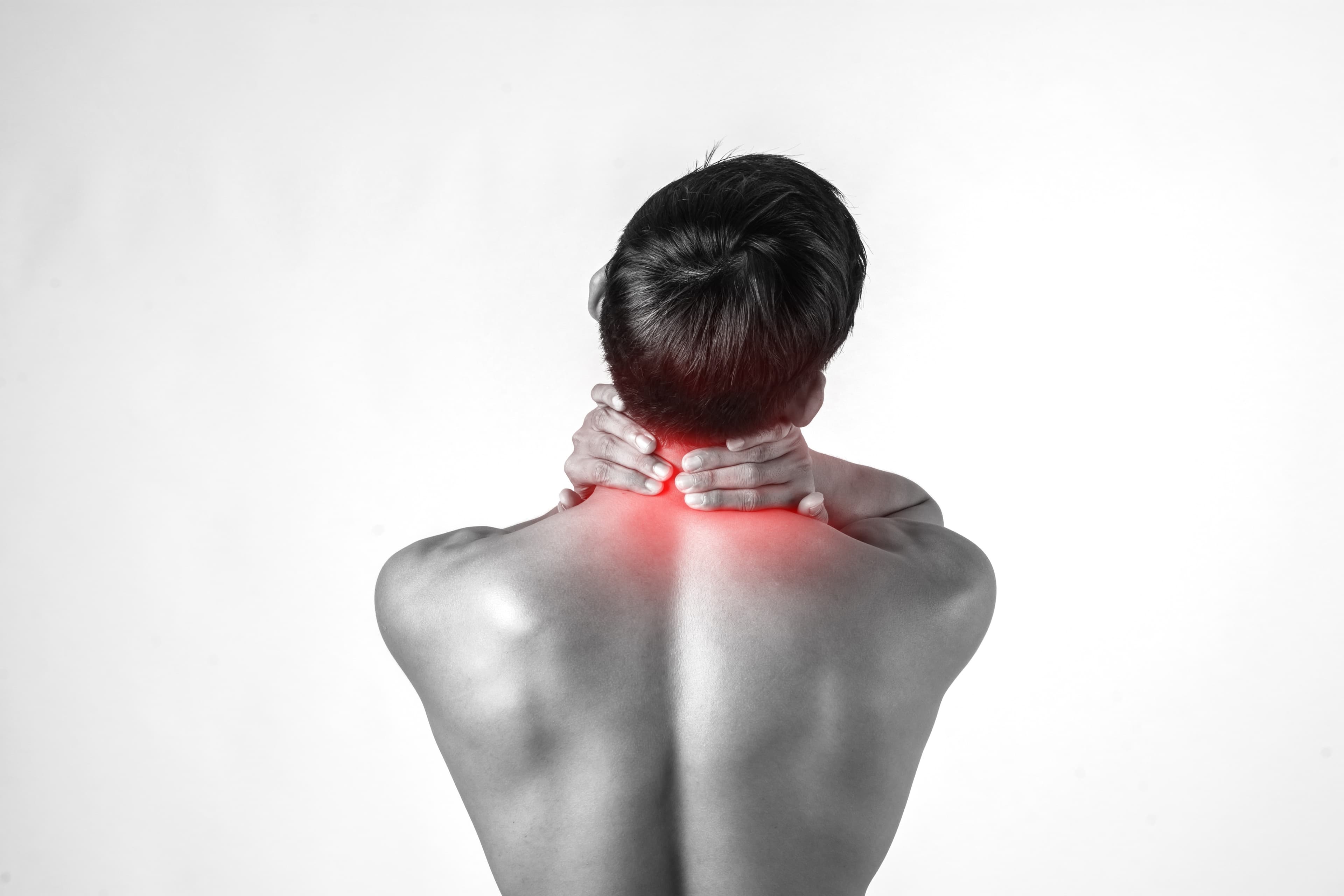
The spine contains both the spinal cord and the nerve roots that branch off from it, exiting through narrow channels between vertebrae to supply the arms and legs. When those channels narrow — from disc degeneration, arthritis, bone spurs, or thickened ligaments — the nerves get compressed.
Compressed spinal nerves don't behave quietly. They cause pain, numbness, weakness, and — in the most disabling form — an inability to walk more than a short distance without severe leg pain or weakness forcing you to stop. This last pattern, called neurogenic claudication, is the classic presentation of lumbar spinal stenosis.
When conservative management (physiotherapy, injections, medication) fails to provide adequate relief, spinal decompression surgery creates space for the nerves by removing the tissue that is compressing them. This guide explains what that surgery involves, which type is appropriate when, and what recovery looks like.
What Causes the Compression?
Spinal stenosis — narrowing of the spinal canal or the nerve exit channels — develops when any of the following structures enlarge or encroach:
Ligamentum flavum hypertrophy: The thick elastic ligament at the back of the spinal canal thickens with age and disc degeneration, bulging into the canal from behind.
Facet joint arthritis and osteophytes: The facet joints degenerate and develop bony spurs that narrow the canal and foramina (nerve exit channels) from the sides and back.
Disc bulging or herniation: The disc bulges or herniates into the canal from the front.
Degenerative spondylolisthesis: One vertebra slips forward over the one below, compressing the nerve roots in the canal between them.
The most common location is the lumbar spine (lower back) — causing leg symptoms. Cervical stenosis (neck) causes arm symptoms and, in more severe cases, spinal cord compression with both arm and leg effects (myelopathy).
The Decision to Operate
Surgery for spinal stenosis is appropriate when:
- Symptoms significantly limit daily activities and quality of life
- Symptoms have persisted despite genuine conservative management — physiotherapy for at least 3 months, injections tried and failed
- There is objective evidence of nerve compression on MRI or CT that correlates with the symptoms clinically
- Progressive neurological deficit: worsening weakness, expanding sensory loss
- Cauda equina syndrome (bladder/bowel involvement from severe central canal stenosis): this is an emergency
The correlation between imaging findings and symptoms is critical. MRI may show significant stenosis in a patient who is functioning well — surgery on the basis of imaging alone, without corresponding clinical symptoms, is inappropriate. Equally, patients with obvious symptoms and clear imaging evidence of corresponding compression are good surgical candidates.
The Types of Spinal Decompression Surgery
Laminotomy
Removal of a small portion of the lamina (the bony roof of the spinal canal) on one side, creating a small window of decompression.
Best for: Single-level or focal compression with contained pathology. Less bone removal means less disruption to the posterior spinal structure. Typically minimally invasive.
Laminectomy
Removal of the entire lamina at one or more levels, plus the spinous process and portions of the thickened ligamentum flavum. Creates a wide decompression of the entire canal.
Best for: Multi-level central canal stenosis causing neurogenic claudication. More extensive decompression but removes more stabilising structure. If the facet joints are also partly removed (facetectomy), the surgeon must assess whether spinal stability is maintained.
When is fusion added to laminectomy? If significant spinal instability exists — either pre-existing (spondylolisthesis) or created by the extent of bone removal — fusion (connecting adjacent vertebrae with rods, screws, and bone graft) may be added to the decompression. Adding fusion prevents the deformity or instability that can result from extensive decompression alone.
Foraminotomy
Enlargement of the foramen (the lateral exit channel where nerve roots leave the canal). Used specifically for foraminal stenosis where a nerve root is compressed laterally rather than centrally. Often performed alongside laminotomy or laminectomy.
Laminoplasty
Used in the cervical spine for multi-level cervical myelopathy. Rather than removing the laminae, they are cut on one side and "opened" like a door on a hinge, expanding the canal while keeping most of the posterior structure intact. Preserves more stability than laminectomy in the cervical spine.
Microdiscectomy
Technically a decompression procedure — removing a herniated disc fragment that is compressing a nerve root. Already covered in the MISS blog, but worth noting here for completeness.
The Operation: What Happens
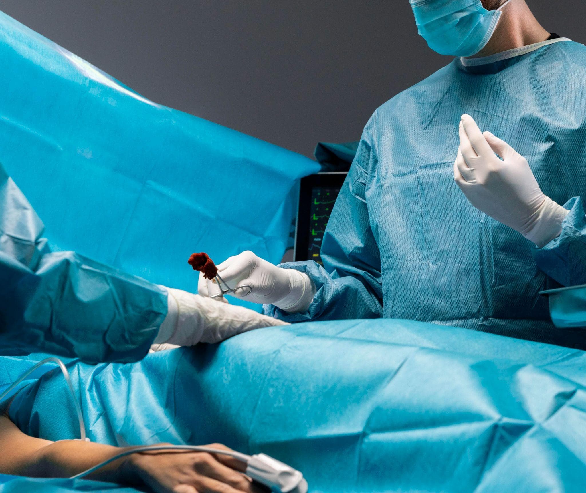
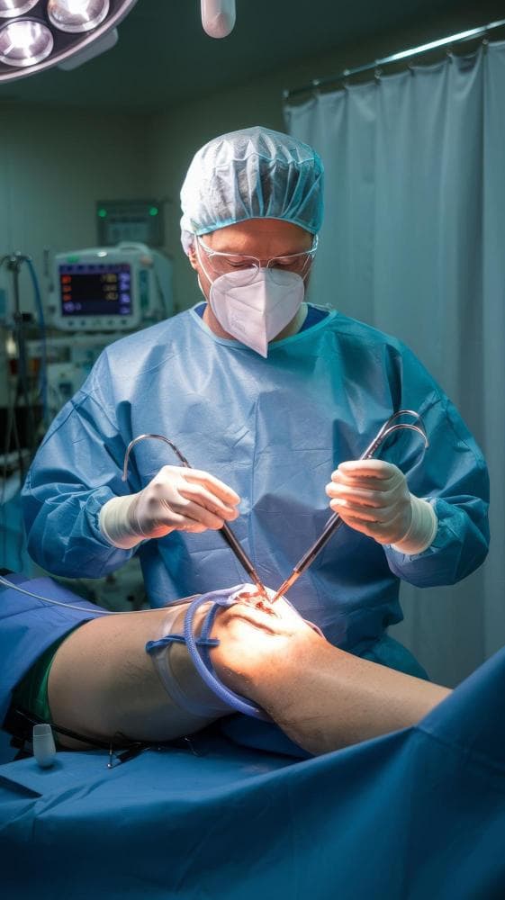
Spinal decompression is typically performed under general anaesthesia, in the prone position (face-down on a specialised frame).
Approach: An incision is made over the affected vertebral level(s) at the back of the spine. The paraspinal muscles are retracted to expose the posterior elements.
Decompression: Using bone rongeurs, a high-speed burr, and Kerrison punches, the surgeon removes the lamina, thickened ligamentum flavum, and osteophytes causing compression. Nerve roots are identified and confirmed to be free of compression before closure.
Minimally invasive variants: Rather than a conventional midline incision, tubular retractor systems allow the same decompression through a small skin incision and a tubular working channel. Less muscle damage, faster recovery, more complex setup.
Fusion (if required): Pedicle screws are placed into the vertebral bodies, connected by rods, and bone graft is placed to fuse the segments.
Closure: Muscles and skin are closed in layers.
Duration: 45–90 minutes for single-level decompression. Multi-level or decompression-plus-fusion cases: 2–4 hours.
Recovery
Single-level laminotomy/microdiscectomy: Often same-day or overnight. Walking the same day or next. Return to desk work: 2–4 weeks. Physical work: 6–8 weeks.
Multi-level laminectomy: 2–4 days in hospital. Walking day 2. Return to normal daily activity: 4–8 weeks. Full recovery: 3–6 months.
Laminectomy with fusion: 3–5 days in hospital. More extensive physiotherapy. Full recovery: 3–12 months depending on complexity.
The relief of leg pain from nerve decompression is often rapid — many patients notice improvement within days of surgery. Back pain recovery is slower and less predictable.
Spine Care at Prakash Hospital, Noida
Dr. Mayank Chauhan evaluates lumbar and cervical spinal stenosis at Prakash Hospital, Sector 33, Noida. Conservative and surgical options are assessed for each patient individually, with MRI review and clinical correlation.
To book a consultation, call the number listed on the website.