Reviewed by Dr. Mayank Chauhan
Bone Cancer and Bone Tumors — Warning Signs, Diagnosis, and Treatment in India
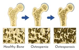
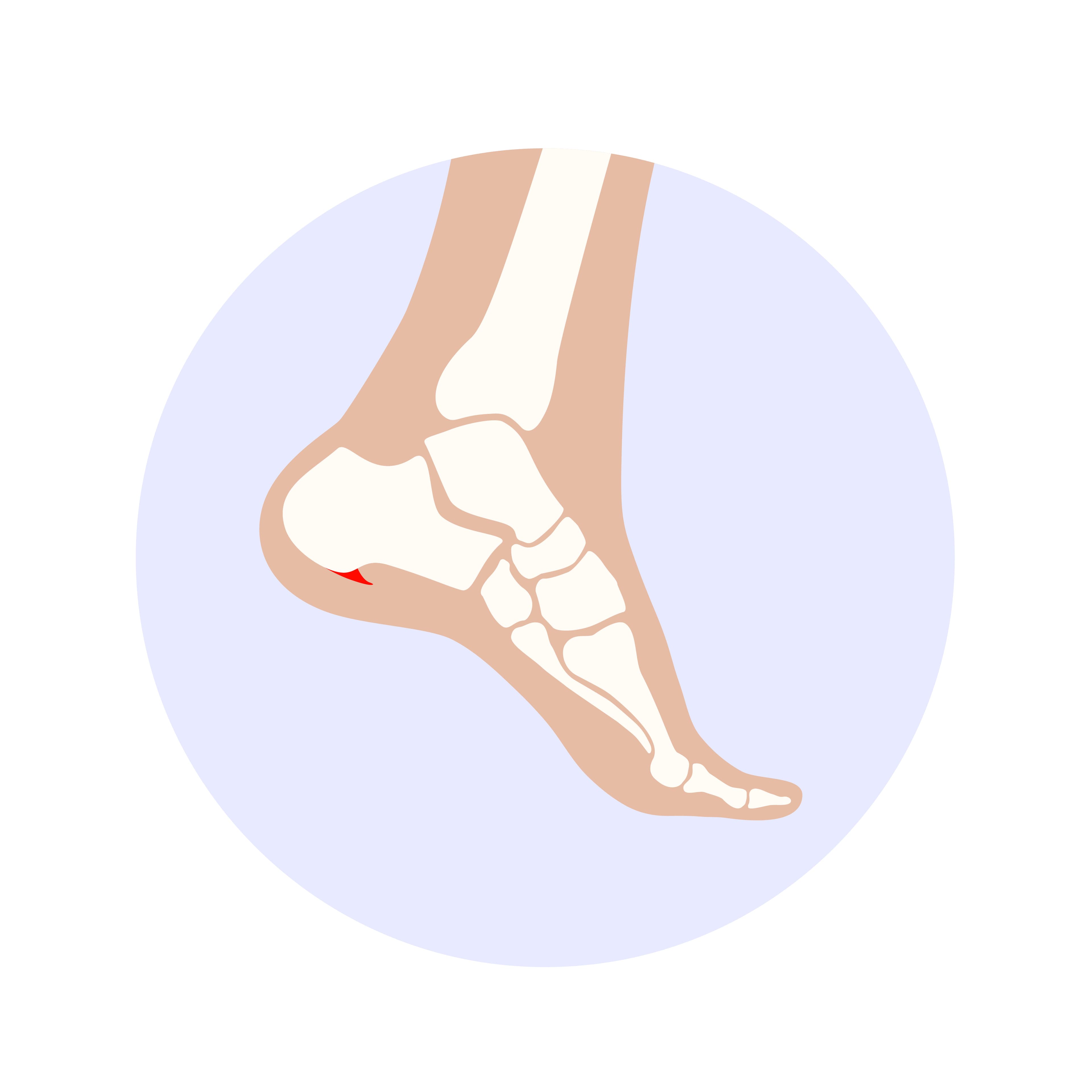
A visual representation of a bone developing Tumor.
Bone pain is common. It has many ordinary causes — muscle strains, fractures from sports, arthritis, the familiar ache of fatigue. Most bone pain resolves with rest, physiotherapy, and time.
But some bone pain doesn't. Some bone pain worsens despite normal recovery. Some swellings appear around bones without a clear history of injury. Some fractures occur from forces too minor to break a healthy bone. These situations require a different kind of evaluation — one focused not on the usual musculoskeletal diagnoses, but on the possibility of bone pathology.
Bone tumors — both benign (non-cancerous) and malignant (cancerous) — are rare compared to soft tissue and organ cancers. Primary bone cancers account for approximately 0.5–1% of all registered malignancies in India, according to data from the National Cancer Registry Programme. But rare doesn't mean unimportant: in young patients particularly, bone tumors can be aggressive, and delayed diagnosis has significant consequences.
Benign Bone Tumors: Common and Usually Not Dangerous
Most bone tumors are benign — they grow locally but do not spread to other organs. Many are discovered incidentally on X-rays taken for other reasons (a sprained ankle, a fall). Many require no treatment.
Osteochondroma (Exostosis)
The most common benign bone tumor. A bony outgrowth covered by a cartilage cap, typically appearing near the growth plate of long bones in adolescents. Most are completely asymptomatic — a painless lump. Treatment is only necessary if it causes symptoms (pain, nerve or vessel compression) or grows unusually large.
Enchondroma
A benign cartilage tumor forming within the bone, typically in the hands and feet. Most are found incidentally on X-ray. Rarely cause problems. Monitored with serial X-rays. Occasionally need biopsy to exclude low-grade malignancy.
Giant Cell Tumor of Bone
Benign but locally aggressive. Occurs most commonly at the ends of long bones near the knee (distal femur, proximal tibia, distal radius). Causes pain and local swelling. Despite being benign, giant cell tumors expand into bone and can cause significant structural damage. Treatment is surgical — curettage (scraping out the tumor) and packing with bone cement or graft.
Bone Cysts (Unicameral and Aneurysmal)
Fluid-filled cavities within bone. Can weaken the bone significantly, causing pathological fracture (fracture through abnormal bone). Treatment ranges from injection to surgical curettage depending on size and location.
Malignant Bone Tumors: Primary Bone Cancer
Osteosarcoma
The most common primary malignant bone tumor in India, particularly in children and adolescents. It arises from osteoblasts (bone-forming cells) and typically occurs during periods of rapid skeletal growth — the second decade of life.
Common sites: Around the knee (distal femur, proximal tibia) — the fastest-growing parts of the skeleton. Also the proximal humerus (upper arm).
Symptoms: Deep, persistent bone pain — initially intermittent, becoming constant. Localised swelling and warmth. Pain characteristically worsens at night and does not respond to rest. Pathological fracture in some cases.
Treatment: Modern treatment combines chemotherapy (neoadjuvant — given before surgery to shrink the tumor and treat microscopic spread) and surgery (limb-salvage resection rather than amputation in most cases, with replacement using prostheses or allografts). Response to chemotherapy is one of the strongest prognostic factors. Post-operative chemotherapy follows. Survival rates with modern protocols: 60–70% five-year survival for localised disease.
Limb-salvage surgery: In many patients, the tumor-bearing segment of bone is removed and replaced with a metal prosthesis or biological reconstruction, preserving the limb and most of its function. This requires highly specialised orthopedic oncology expertise.
Ewing Sarcoma
The second most common primary bone malignancy in children and young adults. Arises from neural crest cells within bone marrow. Can affect any bone but most commonly the femur, pelvis, and ribs.
Symptoms: Localised bone pain, swelling, sometimes fever and elevated inflammatory markers (mimicking infection). The presentation can be easily confused with osteomyelitis (bone infection).
Treatment: Also multimodal — chemotherapy combined with surgery, radiation, or both depending on tumor location and resectability.
Multiple Myeloma
Not a primary bone tumor in the traditional sense — myeloma is a haematological malignancy (cancer of plasma cells in the bone marrow). However, it causes multiple lytic lesions (holes in bone) and is the most common malignancy found in bone in adults, particularly over 50. Presents with bone pain, pathological fractures, and elevated calcium. Managed by haematologists, with orthopedic involvement for fracture stabilisation and cord decompression when needed.
Secondary (Metastatic) Bone Cancer
By far the most common malignant condition affecting bone. When cancers of the breast, lung, prostate, kidney, or thyroid spread to bone — as they frequently do in advanced disease — they produce bone metastases.
Symptoms: Deep, persistent, sometimes severe bone pain at specific sites. Night pain. Pathological fracture. Spinal cord compression from vertebral metastasis.
Treatment: Focuses on pain control, preventing fractures, and addressing spinal involvement. Radiation, systemic oncology treatment, and surgical stabilisation of impending or actual fractures.
Warning Signs That Need Prompt Evaluation
Any of the following bone-related symptoms warrant prompt medical assessment — they are not always cancer, but they need proper evaluation:
- Bone pain that is persistent, progressive, and not explained by trauma or overuse — particularly if it worsens at night
- Pain at rest in a bone — musculoskeletal pain from arthritis or strain typically improves with rest; bone tumor pain often does not
- Swelling or a lump around a bone — firm, fixed, non-tender swelling near a bone is a red flag
- Fracture from minimal or no trauma — breaking a bone from a gentle fall, a cough, or turning in bed suggests the bone may be abnormal (pathological fracture)
- Height loss and severe back pain — in patients over 50, vertebral collapse from metastases or myeloma
- Unexplained weight loss, fever, or night sweats alongside bone pain — suggests systemic malignancy with bone involvement
Diagnosis: The Investigations
X-ray: The first step. Shows bone architecture, any abnormal density changes, periosteal reaction (bone forming around a tumor), and fractures. An experienced radiologist can often characterise a lesion on X-ray.
MRI: The most informative modality for characterising bone and soft tissue extent of a tumor. Shows the marrow, soft tissue involvement, and relationship to neurovascular structures.
CT scan: For precise bony detail and cortical integrity. Also used to stage the disease (check for spread to lungs and other organs).
PET-CT scan: For staging in malignant tumors — detecting distant metastases.
Biopsy: The definitive diagnostic step. A needle biopsy (percutaneous, image-guided) or open surgical biopsy obtains tissue for histopathological analysis. Biopsy should be performed by or in coordination with the team that will perform the definitive surgery — a poorly planned biopsy can compromise subsequent limb-salvage surgery.
Bone scan (isotope scintigraphy): Shows metabolic activity in bone throughout the body. Used to detect multiple lesions and assess disease extent.
Key Points for Indian Patients
- Primary bone cancer is rare but occurs most commonly in the 10–25 age group
- Night pain, persistent unexplained bone pain, and bone swellings in young patients should not be attributed to "growing pains" without proper evaluation
- Limb-salvage surgery has replaced amputation as the standard for most primary bone malignancies at specialist centres in India
- Orthopedic oncology is a subspecialty — management of bone tumors should be at a centre with dedicated oncology infrastructure, including paediatric oncology for children with osteosarcoma
Assessment at Prakash Hospital, Noida
Dr. Mayank Chauhan evaluates bone lesions and provides initial assessment of bone pain with worrying features. Patients with suspected bone tumors are referred appropriately to orthopedic oncology centres for specialist management.
For bone pain evaluation or a second opinion on bone imaging findings, call the number listed on the website.