DEXA Scan — What Is It, Who Needs It, and What the Results Actually Mean

Doctor explaining a scan result to a patient.
Every year, thousands of patients across India receive a DEXA scan report, look at the T-score, compare it to the reference ranges printed at the bottom, and still have no clear idea what the number means for their life.
Does -1.8 mean I'll fracture if I fall? Should I be on medication? Is it going to get worse? What can I actually do about it?
These are not complicated questions, but they rarely get answered in the time available at a radiology appointment. This guide answers all of them.
What a DEXA Scan Is
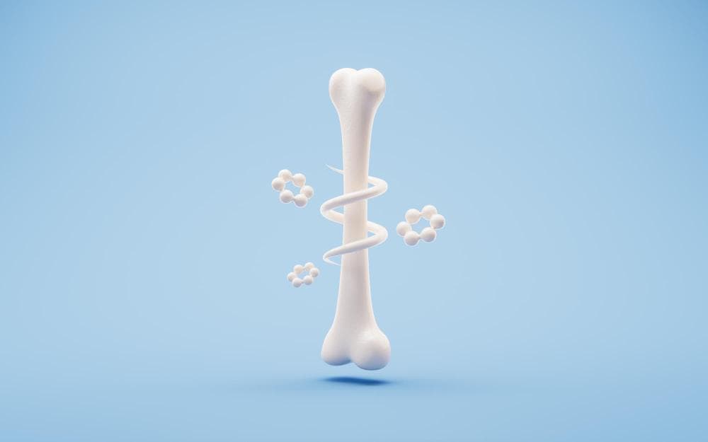
DEXA stands for Dual-Energy X-ray Absorptiometry. It's a low-dose X-ray test that measures the mineral content — primarily calcium — in specific bones, and from that measurement calculates bone mineral density (BMD).
The scan typically takes 15–20 minutes. You lie on a padded table, fully clothed (no metal fasteners, which interfere with imaging). A scanner arm passes slowly over your body. Two X-ray beams of different energy levels are projected through the bone. Because different tissues (soft tissue, fat, bone) absorb these beams differently, the machine can isolate and measure bone mineral content with high precision.
It is painless, non-invasive, and uses radiation doses far lower than a chest X-ray.
The standard DEXA scan measures two sites: the lumbar spine (L1–L4 vertebrae) and the femoral neck (top of the hip bone, just below where the hip ball meets the pelvis). These are the sites where osteoporotic fractures are most likely to occur — and where treatment decisions are made.
Some scans also measure the distal radius (one-third point of the forearm bone) — this is particularly useful in certain situations, such as patients on parathyroid hormone-related medication.
What the T-Score Means
The report will give you several numbers. The most important one for clinical decision-making is the T-score at each site.
The T-score compares your bone mineral density to the average peak bone density of a healthy young adult (typically a woman in her late 20s). The comparison is expressed in standard deviations above or below that reference:
| T-score | Category | Interpretation |
| -1.0 or above | Normal | Bone density is within the healthy range |
| -1.0 to -2.5 | Osteopenia (low bone mass) | Bone density is below average; monitor and address risk factors |
| -2.5 or below | Osteoporosis | Bone density is significantly low; fracture risk is elevated |
These thresholds were established by the WHO and are the same worldwide.
A few important points that are often missed
The lowest site drives the diagnosis. If your spine T-score is -1.5 but your hip T-score is -2.7, you have osteoporosis — based on the lower score.
T-score is not the whole story. Two patients with the same T-score can have very different fracture risks, depending on their age, sex, prior fractures, medications, and other health factors.
Osteopenia does not mean fracture is inevitable. Many people with osteopenia never fracture. Others with relatively less severe osteopenia do fracture, because of other risk factors.
The Z-Score: A Different Comparison
Your report will also show a Z-score. This compares your bone density to other people of your same age, sex, and ethnicity.
The Z-score is clinically useful in a different way from the T-score: it tells you whether your bone density is where it should be for your age, not just compared to a young adult.
A low Z-score (below -2.0) in an older adult suggests that bone loss is happening faster than normal ageing would explain — which should trigger a search for secondary causes: corticosteroid use, hyperparathyroidism, coeliac disease, hyperthyroidism, premature menopause, or other conditions that accelerate bone loss.
If your Z-score is low, don't ignore it. It's asking a question about why.
How to Read Your Report
Your DEXA report will typically include:
- BMD values at each site (spine L1–L4, femoral neck, total hip), expressed as grams per square centimetre
- T-score at each site
- Z-score at each site
- A graph showing where your score sits relative to the reference population (often colour-coded: green = normal, yellow = osteopenia, red = osteoporosis)
The site with the lowest T-score is the one that determines your classification. Look across all sites and find the lowest number — that's your diagnostic category.
If you've had a previous DEXA scan, the report may also show rate of change — how much bone density has changed since the last scan. Meaningful loss (more than 3–5% at the spine or hip over 1–2 years) may prompt treatment changes.
Who Should Get a DEXA Scan?
Not everyone needs one. But these groups have a clear indication:
Women aged 65 and older: Universal screening recommendation across most guidelines. Bone loss after menopause is universal; screening identifies those whose loss has crossed into osteoporosis territory.
Postmenopausal women under 65 with risk factors: Family history of hip fracture, prior fragility fracture, early menopause (before 45), low body weight (BMI below 19), smoking, excessive alcohol, long-term corticosteroid use.
Men aged 70 and older: Men also develop osteoporosis, particularly after 70. Screening is recommended at this age.
Anyone with a fragility fracture: A fracture from a fall from standing height or less — regardless of age or sex — should trigger bone density assessment. This fracture is telling you something about bone quality.
Patients on long-term corticosteroids: Steroid-induced osteoporosis is common and underdiagnosed. Any patient expected to be on systemic steroids for 3 months or more should have a baseline DEXA.
Patients with conditions associated with bone loss: Rheumatoid arthritis, coeliac disease, inflammatory bowel disease, hyperparathyroidism, hyperthyroidism, chronic kidney disease, liver disease.
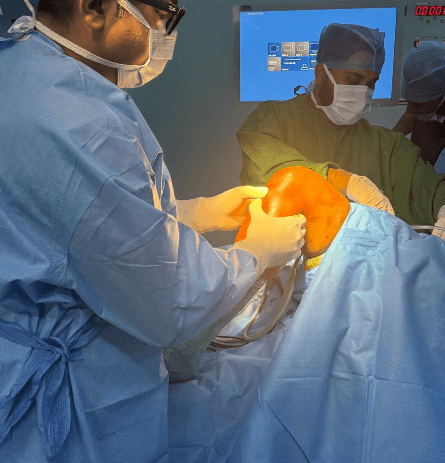
Patients before orthopedic surgery: At Prakash Hospital Noida, bone quality assessment is part of pre-operative evaluation for joint replacement. Operating on very osteoporotic bone requires planning adjustments.
How Often Should You Repeat It?
Once a baseline scan has been done, the frequency of repeats depends on your results and risk category:
- Normal bone density, no major risk factors: Every 10–15 years is sufficient
- Osteopenia with moderate risk: Every 2–3 years
- Osteoporosis under treatment: Every 1–2 years (to monitor treatment response)
- After a fragility fracture: Baseline scan urgently, then annually to monitor
If a repeat scan is done too soon (less than 12–18 months after the previous one), the change in density may be within the machine's margin of error — making it difficult to distinguish real change from measurement variation.
What Happens If Your Results Are Abnormal?
Osteopenia: In most cases, lifestyle modification is the primary response — calcium, vitamin D, exercise, smoking cessation. A repeat scan in 1–2 years tracks whether measures are working. Medication may be appropriate for patients with high calculated fracture risk (assessed using FRAX tool) even with osteopenia.
Osteoporosis: Medication is usually recommended alongside lifestyle measures. The most commonly used drugs are bisphosphonates (alendronate, risedronate, zoledronic acid). The decision about which medication, in what form, for how long, requires a specialist assessment.
High Z-score (more loss than expected for age): Investigation for secondary causes before starting treatment.
DEXA Scanning at Prakash Hospital, Noida
Prakash Hospital at Sector 33, Noida, has on-site DEXA bone density scanning available. Results are interpreted in the context of a clinical evaluation by Dr. Mayank Chauhan — not just handed over as a number.
If you have risk factors for osteoporosis, have had a previous fracture, or are approaching or past menopause and haven't had a bone density check, a DEXA scan is a reasonable next step.
To book a consultation, call the number listed on the website.