Ankle Sprain - Grade 1, 2, And 3: How to Treat It Properly and Avoid Chronic Instability

A young man is kneeling and holding his right ankle, and has an anxious look on his face. In the background, there is an athletic field all around. The skin surrounding his ankle is a little reddish, which indicates pain.
Ankle sprains are the single most common sports injury worldwide — and the most consistently undertreated. In Noida's gyms, cricket grounds, badminton courts, and running tracks, a rolled ankle is met with the same predictable response: a few minutes of limping, some application of pain-relief spray, a compression bandage from the first aid kit, and a return to activity within a day or two.
Sometimes this works out fine. Grade 1 sprains genuinely do resolve with basic management. But Grade 2 and 3 sprains — the more significant tears that produce real structural ligament damage — routinely receive the same inadequate treatment as Grade 1 sprains. The result is predictable: the ligament never fully heals, the ankle becomes chronically unstable, and the patient has the first of what will be many repeat sprains.
Chronic ankle instability — the consequence of inadequately managed ankle sprains — is one of the most common orthopedic problems in active adults in India. And it is largely preventable.
Anatomy of the Ankle — Why the Lateral Side Is Vulnerable
The ankle joint connects the tibia and fibula (the two leg bones) to the talus (the ankle bone). Three groups of ligaments provide stability:
Lateral (outer) ligaments — injured in the vast majority of ankle sprains:
- ATFL (Anterior Talofibular Ligament): The most commonly injured ankle ligament — connects the fibula to the talus, preventing anterior sliding of the talus. Injured first in most ankle sprains.
- CFL (Calcaneofibular Ligament): Connects the fibula to the heel bone (calcaneus). Injured in more severe sprains — alongside the ATFL.
- PTFL (Posterior Talofibular Ligament): The strongest lateral ligament; injured only in the most severe sprains or dislocations.
Medial (inner) ligaments (deltoid ligament): Much stronger than the lateral complex; injured far less commonly — typically from eversion (outward rolling) injuries. When the deltoid ligament tears, the force required is significant, and associated injuries (fractures) are common.
Syndesmotic ligaments: Connect the tibia and fibula above the ankle joint. Injured in high ankle sprains — from forced external rotation — which are more common in contact sports and skiing. Significantly more serious than lateral sprains; often require longer recovery.
Why the lateral side is more vulnerable:
The lateral ligaments are mechanically weaker than the medial deltoid complex. The anatomy of the foot and ankle naturally biases toward inversion (rolling inward) — the most common ankle sprain mechanism. Approximately 85% of all ankle sprains are inversion-type injuries affecting the lateral ligament complex.
How Ankle Sprains Happen
The classic mechanism: the foot lands in an inverted position (sole turned inward, top of the foot turning outward) while the leg remains upright. This positions the lateral ligaments under tension — and if the force exceeds the ligament's capacity, tearing occurs.
Common scenarios in Noida:
- Landing awkwardly from a jump in badminton, basketball, or cricket fielding
- Stepping off a kerb or into a pothole
- Running on uneven road or park surfaces
- Sudden direction changes in football, futsal, or kabaddi
- Walking in hawai chappals (rubber slippers) that provide no ankle support — extremely common
- Gym exercises involving lateral movements or plyometrics without adequate footwear
High-risk footwear in India:
Hawai chappals and other minimal footwear with no lateral ankle support are a significant risk factor for ankle sprains in the urban Indian population — not just in sport, but in everyday walking. The transition from sandals to athletic footwear for sports should be a standard recommendation.
Grading the Injury — The Critical First Step
The management of an ankle sprain is fundamentally different depending on its grade. Treating a Grade 3 tear the same way as a Grade 1 sprain — as commonly happens — leads to inadequate healing and chronic instability.
Grade 1 (Mild Sprain)
Ligament status: Fibres stretched but structurally intact. No macroscopic tearing.
Clinical picture:
- Mild tenderness over the ATFL (the soft spot just in front of the fibula)
- Mild swelling — localised, not dramatic
- Able to bear weight and walk, though with some discomfort
- No clinical instability on examination (the ankle feels stable)
Recovery: 5–14 days.
Treatment: RICE protocol — Rest (avoid aggravating activity), Ice (15–20 minutes every 2 hours for 48 hours), Compression (elastic bandage), Elevation (leg raised above heart level). Paracetamol or NSAIDs for pain. Resume activity progressively as pain allows. Proprioception exercises from Day 3–5 onwards.
Grade 2 (Moderate / Partial Tear)
Ligament status: Partial tearing of the ATFL — the ligament is partially disrupted. In more severe Grade 2 injuries, the CFL is also partially involved.
Clinical picture:
- Moderate to significant pain over the lateral ankle
- Significant swelling — often extending toward the foot; bruising (ecchymosis) appears within 24 hours
- Difficulty bearing weight — may be able to walk with a limp, but walking is clearly painful
- Some clinical instability on stress testing — the ankle shows more laxity than the other side, but the endpoint is firm (there is still some structural integrity)
Recovery: 3–6 weeks.
Treatment: This is where many patients in India self-treat inadequately. Grade 2 sprains need:
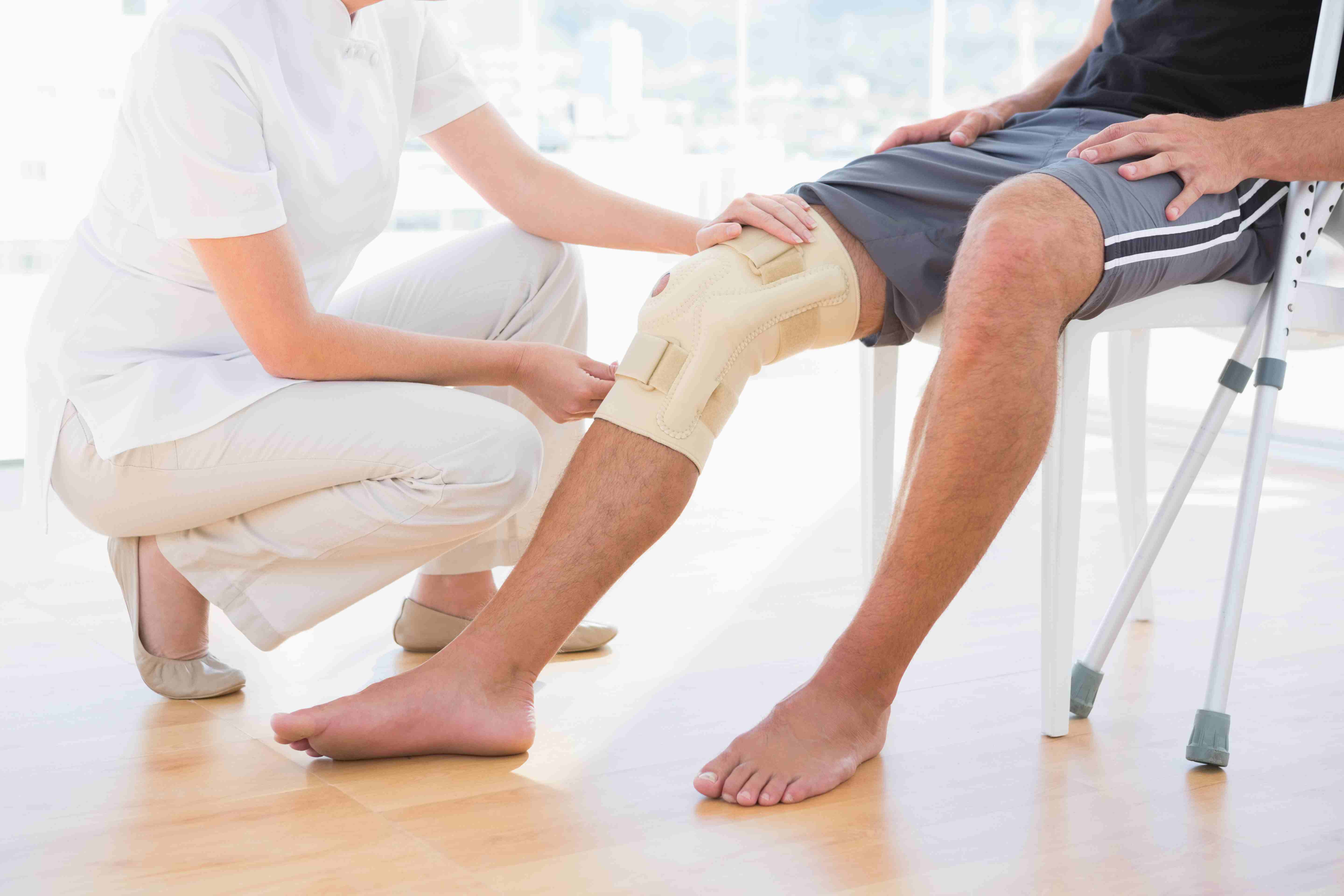
- Protected weight-bearing — a lace-up ankle brace or semi-rigid boot for 1–2 weeks to protect the partially torn ligament while it heals in a shortened position
- Crutches for the first 3–5 days if walking is very painful
- RICE aggressively in the first 48–72 hours
- NSAIDs for 5–7 days for pain and inflammation
- Physiotherapy from Week 2 — range-of-motion exercises progressing to proprioception and strengthening
- No return to sport until proprioception is restored and strength symmetry is confirmed (at least 4–6 weeks)
Grade 3 (Severe / Complete Tear)
Ligament status: Complete rupture of the ATFL and typically the CFL as well. The lateral ankle is structurally unstable.
Clinical picture:
- Severe pain at the moment of injury — often more than expected in a "rolled ankle."
- Rapid, dramatic swelling — haemarthrosis (blood in the ankle joint) may be present
- Inability to bear weight in many cases
- Significant clinical instability — the ankle has clear laxity with a soft endpoint on stress testing
- Bruising that develops over 24–48 hours and extends toward the foot and around the heel
Recovery: 8–12 weeks minimum for ligament healing; return to sport with full training in 3–4 months.
Treatment:
- X-ray to exclude fractures — avulsion fractures of the fibula tip are common with Grade 3 sprains; the Ottawa Ankle Rules guide when X-ray is necessary
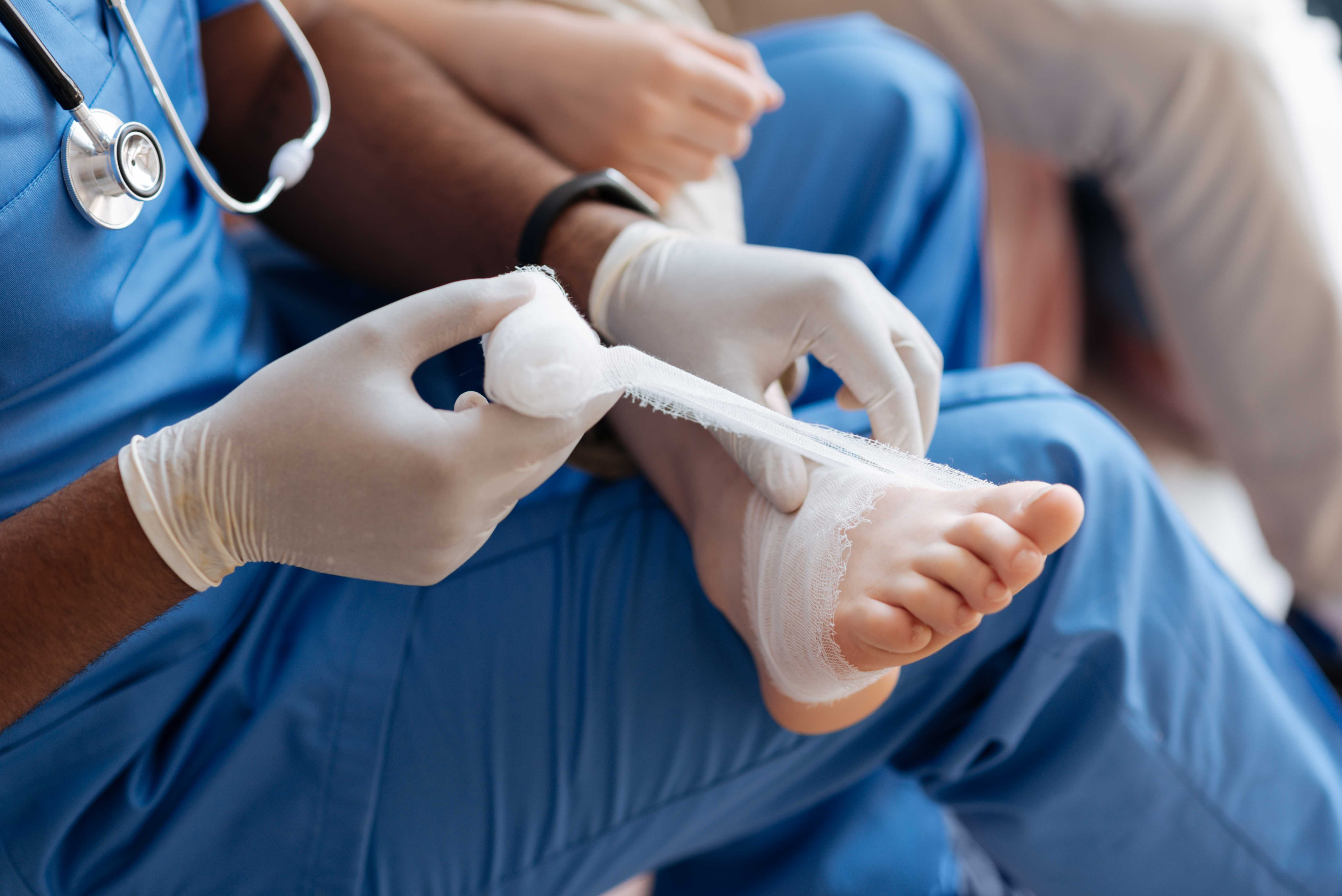
- Immobilisation in a boot (not just a bandage) for 2–3 weeks to allow healing with the ligament ends in approximation
- Crutches for weight-bearing management in the acute phase
- Progressive physiotherapy: range of motion → proprioception → strengthening → sport-specific
- A surgeon's assessment is appropriate to: confirm the diagnosis with MRI (in cases where the clinical picture is severe), exclude associated injuries (osteochondral lesions, peroneal tendon tears), and plan rehabilitation
Surgery for acute Grade 3 sprains:
Most evidence does not support primary surgical repair for acute Grade 3 lateral ankle sprains in the general population — conservative management produces equivalent functional outcomes with lower complication risk. Surgery is reserved for cases of chronic instability that have failed conservative management.
The Ottawa Ankle Rules — When to Get an X-ray
The Ottawa Ankle Rules are a validated clinical decision tool that helps determine when an ankle X-ray is necessary to rule out fracture. They are based on clinical findings:
X-ray is recommended if:
- Bony tenderness at the tip or posterior edge of either malleolus (the bony bumps on either side of the ankle)
- Bony tenderness over the navicular bone (inner midfoot) or the base of the fifth metatarsal (outer midfoot)
- Inability to bear weight (4 steps) immediately after the injury AND at the time of examination
The Ottawa Rules are 95–100% sensitive for clinically significant ankle fractures. If none of these criteria are present, a fracture is very unlikely, and an X-ray can be avoided.
Why this matters:
Many patients in Noida go directly to X-ray after any ankle injury — and then, when the X-ray is normal, are told "no fracture — it's just a sprain" without assessment of the ligament injury severity. An ankle X-ray being normal does not mean the injury is minor. Grade 3 ligament tears have completely normal X-rays.
Physiotherapy for Ankle Sprains — The Crucial Component
Physiotherapy after an ankle sprain is not simply "rest until better." A structured rehabilitation programme — addressing all components of ankle function — determines whether the sprain heals completely or contributes to chronic instability.
Phase 1 — Acute Management (Days 1–5)
Goals: Reduce swelling and pain, protect the healing ligament, begin early movement.
- RICE/POLICE protocol
- Ankle pumps (gentle movement that activates the calf muscle pump and reduces swelling)
- Non-weight-bearing or partial weight-bearing as pain allows
- Compression bandage or brace
Phase 2 — Restoration of Range of Motion (Days 5–14)
Goals: Restore full ankle range of motion.
- Alphabet exercises (tracing the alphabet with the toes — moves the ankle through all ranges)
- Ankle circles
- Calf stretches
- Weight-bearing exercises as tolerated
Phase 3 — Proprioception Training (Weeks 2–6)
This is the most important and most neglected phase. Proprioception is the joint's ability to sense its own position and communicate this to the brain — triggering automatic protective muscle contraction before the ankle rolls.
After a ligament tear, proprioceptive nerve endings in the ligament are disrupted — even after the structural healing is complete, the joint's "alarm system" is impaired. Without specific proprioception retraining, the ankle is vulnerable to repeat sprains even after the ligament has healed.
Proprioception exercises:
- Single-leg standing (balance on the injured ankle with eyes open, then closed)
- Balance board/wobble board exercises
- Bosu ball standing
- Single-leg squat and landing
The degree of proprioceptive recovery is the most important determinant of whether the ankle will roll again.
Phase 4 — Strength and Return to Sport (Weeks 4–12)
- Peroneal muscle strengthening (the evertor muscles on the outer calf — the primary dynamic stabilisers of the lateral ankle)
- Calf raise exercises (standing and single-leg)
- Progressive agility training: straight-line running → cutting → sport-specific movements
- Return to sport only when: full range of motion, 90%+ strength symmetry, proprioception tests passed, no pain with sport-specific movements
Chronic Ankle Instability — When a Sprain Becomes a Long-Term Problem
Chronic ankle instability (CAI) is the condition that develops when an ankle sprain is inadequately treated — or when repeated sprains damage the lateral ligament complex beyond its capacity to heal.
What CAI feels like:
- The ankle "gives way" with activities that previously felt safe — walking on uneven ground, descending stairs, sports
- Persistent pain and swelling after activity
- A general sense that the ankle is unreliable
- Loss of confidence in the ankle during sports or any dynamic activity
Prevalence:
Studies consistently show that approximately 30–40% of ankle sprain patients develop CAI — mostly from Grade 2 and 3 injuries that were not adequately rehabilitated.
Management of CAI:
- Intensive proprioceptive rehabilitation — the most important intervention for functional instability
- Ankle foot orthosis (brace) for sport
- Peroneal strengthening
- Surgical stabilisation — when conservative management fails: the Broström-Gould procedure (repair and reinforcement of the ATFL and CFL) is the gold standard, producing 85–95% good outcomes
Ankle Sprain and Chronic Instability Management in Noida — Dr. Mayank Chauhan
Dr. Mayank Chauhan, Senior Orthopedic Surgeon at Prakash Hospital, Sector 33, Noida, evaluates ankle injuries — acute sprains and chronic instability — providing accurate grading, appropriate imaging when indicated, and a structured rehabilitation plan matched to the grade of injury.
For patients in Noida and Greater Noida who have had a significant ankle sprain, or who are dealing with an ankle that keeps giving way months or years after an initial injury, proper evaluation will clarify the diagnosis and the management path. To book a consultation, call the number listed on the website.
The Bottom Line
Ankle sprains are common — but they are not trivial, and they are not all the same. Grade 1 sprains heal quickly with basic management. Grade 2 and 3 sprains require protected healing, structured physiotherapy, and specific proprioception retraining to prevent the chronic instability that affects 30–40% of inadequately managed cases. The right treatment — matched to the specific grade — produces complete recovery. The wrong treatment produces a repeat injury.
To consult Dr. Mayank Chauhan, Senior Orthopedic Surgeon in Noida, call the number listed on the website.