Avascular Necrosis of the Hip — Causes, Stages, and All Treatment Options
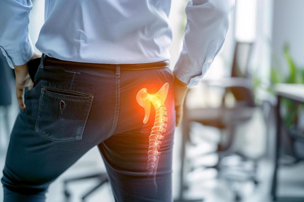
A man is standing with one hand on his hip, a glowing illustration of the hip bone is overlaid, indicating pain or an issue in that area.
Avascular necrosis of the hip is one of the most feared diagnoses in orthopedic medicine — and one of the most misunderstood.
It's not arthritis, though it eventually looks a lot like it. It's not a fracture, though the hip can collapse as if it were. It's a condition in which the blood supply to the femoral head (the ball of the hip joint) is cut off or reduced, causing bone tissue to die. Without blood, bone cannot regenerate. The structural integrity of the femoral head gradually fails. The smooth ball that should glide frictionlessly in its socket becomes irregular, collapsed, and painful.
In India, AVN of the hip has a particularly important profile. The steroid use associated with COVID-19 treatment between 2020 and 2022 triggered a significant wave of post-COVID AVN cases — many of them in young patients, many of them bilateral. A retrospective study of 212 hips with post-COVID AVN found a mean patient age of just 36.8 years, with 79.7% presenting with bilateral disease. These are patients in the middle of working life, often with young families, facing the prospect of hip replacement decades earlier than anyone expected.
Understanding AVN — its causes, how it progresses, and when each treatment is appropriate — matters more in India today than it has in a very long time.
What Causes Avascular Necrosis of the Hip?
AVN occurs when blood flow to the femoral head is disrupted. Several mechanisms can cause this:
Corticosteroid use: The most common non-traumatic cause in India. High-dose or prolonged corticosteroid therapy — used for autoimmune diseases, inflammatory conditions, organ transplant rejection, and extensively during the COVID-19 pandemic — is directly associated with AVN. Steroids appear to cause fat deposits in small blood vessels supplying the femoral head, reducing blood flow. The risk increases with cumulative dose and duration.
Traumatic causes: Hip dislocations and femoral neck fractures can damage the blood vessels that supply the femoral head directly. This is one reason femoral neck fractures in younger adults are treated urgently.
Excessive alcohol consumption: The second most common non-traumatic cause, and one with significant prevalence in India. Alcohol increases fat levels in the blood and causes fat emboli that block the vessels supplying the femoral head. The risk is dose-dependent.
Sickle cell disease: More prevalent in tribal populations of Central and Western India. The abnormal, sickle-shaped red blood cells obstruct small vessels and are a major cause of AVN in these populations.
Medical conditions: Systemic lupus erythematosus (SLE), coagulation disorders, and Gaucher's disease all increase AVN risk.
Idiopathic: In some cases, no clear cause is identified. The blood supply is disrupted for reasons that cannot be definitively established.
Why Is Early Diagnosis So Important?
AVN is a progressive disease. It moves through defined stages, and the options available — and the prognosis — change at each stage.
In the earliest stages, the bone is weakened but the femoral head has not yet collapsed. If diagnosed here, there are surgical options (core decompression, bone grafting) that can potentially preserve the natural hip joint and delay or prevent the need for hip replacement.
By the time the femoral head has collapsed, the cartilage above it is destroyed, the joint is irregular, and the symptoms are those of severe arthritis. At this stage, preserving the natural joint is no longer possible. Hip replacement becomes the appropriate option.
The tragedy of AVN is that the early stages are often silent. Many patients have no pain in Stage I or Stage II. By the time pain brings them to a doctor, they may already be in Stage III or IV.
This is why any patient with significant corticosteroid exposure, a history of alcohol misuse, or post-COVID treatment history who develops hip or groin pain should have an MRI early — not just an X-ray. MRI detects AVN far earlier than X-ray.
The Ficat and Arlet Staging System
The most widely used classification for AVN of the hip:
Stage I
X-rays appear entirely normal. The diagnosis can only be made on MRI, which shows early bone marrow changes. The femoral head is still intact. No collapse has occurred.
Prognosis without treatment: Most Stage I cases progress. The rate of progression depends on the size of the involved area (the "lesion") and the underlying cause.
Treatment options: Conservative management (activity modification, protected weight-bearing, NSAIDs) is occasionally used but rarely prevents progression. Core decompression is the most evidence-based surgical intervention at this stage.
Stage II
Changes now visible on X-ray — sclerotic (denser) or cystic (lucent) areas in the femoral head, but no collapse yet. MRI shows more extensive involvement.
Prognosis: Still an opportunity to preserve the hip joint, but the window is narrowing.
Treatment options: Core decompression, with or without bone grafting or biological agents (platelet-rich plasma, stem cell augmentation). Vascularized fibular graft is a complex option for young patients with large lesions.
Stage III
The subchondral bone (the thin bone just beneath the cartilage) fractures. On X-ray or CT, this appears as a "crescent sign." The femoral head begins to lose its spherical shape. This is the pivotal stage.
Prognosis: Once the crescent sign is visible, collapse is likely imminent without intervention. Preserving the natural joint is still technically possible but outcomes of joint-preserving surgery are much less reliable at this stage.
Treatment options: For young patients with small lesions, joint-preserving surgery may still be attempted. For most patients, the discussion moves toward hip replacement — osteotomy (repositioning the femoral head to place healthy bone in the weight-bearing zone) in select younger patients, or total hip replacement.
Stage IV
The femoral head has collapsed. The smooth spherical shape is lost. The cartilage above the collapsed bone is damaged. The joint now shows the appearance of advanced arthritis on X-ray.
Treatment: Total hip replacement. There is no realistic alternative at this stage that provides reliable pain relief and restored function. The results of hip replacement for AVN are generally very good.
Treatment in Detail
Core Decompression
The most common surgery for early-stage AVN. One or more small channels are drilled through the femoral neck into the area of bone death. This serves two purposes: it physically relieves the raised pressure within the bone (which is believed to contribute to pain and further vascular compromise), and it creates a pathway for new blood vessels to grow into the area.
Studies show core decompression is most effective in Stage I AVN with smaller lesions, where success rates (preventing progression to collapse) can reach 70–80%. In Stage II, outcomes are more variable. By Stage III, core decompression alone rarely prevents collapse.
Bone Grafting
Structural or vascularized bone grafts can be combined with core decompression to provide support for the weakened subchondral bone. Vascularized fibular grafting — a complex procedure involving transplanting a segment of the fibula bone with its blood supply into the femoral head — is reserved for young patients with large lesions, usually performed at specialized centres.
Osteotomy
Repositions the femoral head so that the healthy (non-necrotic) bone takes the load in the weight-bearing position. Technically demanding and appropriate only for specific lesion locations and younger patients. Used less commonly now than hip replacement in most settings.
Total Hip Replacement
The definitive treatment for Stage III–IV AVN. The damaged femoral head and the socket are replaced with metal and plastic components. This eliminates the pain, restores function, and provides very reliable long-term results.
Hip replacement for AVN in younger patients is a particular consideration. Because the patients may be in their 30s or 40s, implant longevity matters more, and the possibility of revision surgery during their lifetime must be discussed. Modern implants — particularly ceramic-on-ceramic bearing surfaces — are designed for younger, more active patients.
Post-COVID AVN: What Indian Patients Need to Know
The COVID-19 pandemic created a cohort of AVN patients that India's orthopedic community had not seen before: young, often previously healthy individuals, many presenting with bilateral hip AVN within 6–18 months of COVID-19 treatment involving high-dose corticosteroids.
A 2025 study published in the journal Cureus, analysing 118 patients (212 hips) with post-COVID AVN, found that patients presented at a mean age of 36.8 years. The majority had bilateral disease (80%). At 2-year follow-up, a significant proportion required hip replacement.
The message for anyone who received high-dose corticosteroids during COVID-19 treatment and has since developed hip, groin, or thigh pain: do not delay evaluation. An MRI is the appropriate first investigation, not just an X-ray.
When to See an Orthopedic Surgeon
Seek evaluation if:
- You develop hip or groin pain, particularly with activity or weight-bearing
- You have a history of corticosteroid use (for any reason) and develop hip pain
- You had COVID-19 treatment involving steroids
- You have a history of significant alcohol use and develop hip pain
- You have been diagnosed with sickle cell disease, SLE, or other AVN-associated conditions and develop hip symptoms
Early diagnosis is the most important factor in preserving options. Waiting until the pain is severe often means waiting until joint preservation is no longer possible.
AVN Hip Treatment at Prakash Hospital, Noida
Dr. Mayank Chauhan at Prakash Hospital, Sector 33, Noida, has extensive experience in the management of avascular necrosis — from early-stage core decompression to total hip arthroplasty for advanced disease. He evaluates each patient with a careful assessment of staging, patient age, lesion characteristics, and underlying cause before recommending treatment.
Prakash Hospital serves patients from across Noida, Greater Noida, and the Delhi NCR region.
To book a consultation, call the number listed on the website.